 Movie
Movie Controller


+ Open data

- Basic information
| Entry | Database: PDB / ID: 1qst | |||||||||
|---|---|---|---|---|---|---|---|---|---|---|
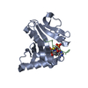
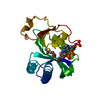
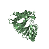
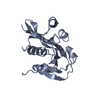
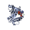
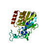
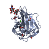
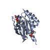
| Title | CRYSTAL STRUCTURE OF TETRAHYMENA GCN5 | |||||||||
 Components Components | TGCN5 HISTONE ACETYL TRANSFERASE | |||||||||
 Keywords Keywords |  TRANSFERASE / HISTONE ACETYLTRANSFERASE / GCN5-RELATED N-ACETYLTRANSFERASE / COA BINDING PROTEIN TRANSFERASE / HISTONE ACETYLTRANSFERASE / GCN5-RELATED N-ACETYLTRANSFERASE / COA BINDING PROTEIN | |||||||||
| Function / homology |  Function and homology informationhistone acetyltransferase activity / histone acetyltransferase / nucleus Function and homology informationhistone acetyltransferase activity / histone acetyltransferase / nucleusSimilarity search - Function | |||||||||
| Biological species |   Tetrahymena thermophila (eukaryote) Tetrahymena thermophila (eukaryote) | |||||||||
| Method | X-RAY DIFFRACTION / SYNCHROTRON / Resolution: 1.7 Å | |||||||||
 Authors Authors | Rojas, J.R. / Trievel, R.C. / Zhou, J. / Mo, Y. / Li, X. / Berger, S.L. / David Allis, C. / Marmorstein, R. | |||||||||
 Citation Citation | Journal: Nature / Year: 1999 Title: Structure of Tetrahymena GCN5 bound to coenzyme A and a histone H3 peptide. Authors: Rojas, J.R. / Trievel, R.C. / Zhou, J. / Mo, Y. / Li, X. / Berger, S.L. / Allis, C.D. / Marmorstein, R. | |||||||||
| History |
|
- Structure visualization
| Structure viewer | Molecule: MolmilJmol/JSmol |
|---|
- Downloads & links
-Download
| PDBx/mmCIF format | 1qst.cif.gz | 47.1 KB | Display | PDBx/mmCIF format |
|---|---|---|---|---|
| PDB format | pdb1qst.ent.gz | 33.6 KB | Display | PDB format |
| PDBx/mmJSON format | 1qst.json.gz | Tree view | PDBx/mmJSON format | |
| Others |  Other downloads Other downloads |
-Validation report
| Arichive directory | https://data.pdbj.org/pub/pdb/validation_reports/qs/1qstftp://data.pdbj.org/pub/pdb/validation_reports/qs/1qst | HTTPS FTP |
|---|
-Related structure data


-Links
 PDBj
PDBj


- Assembly
| Deposited unit | 
| ||||||||
|---|---|---|---|---|---|---|---|---|---|
| 1 |
| ||||||||
| Unit cell |
|
-Components
| #1: Protein | Mass: 19130.256 Da / Num. of mol.: 1 Source method: isolated from a genetically manipulated source Source: (gene. exp.) Tetrahymena thermophila (eukaryote) / Plasmid: PRSET A / Production host:  Escherichia coli (E. coli) / References: UniProt: Q27198 Escherichia coli (E. coli) / References: UniProt: Q27198 |
|---|---|
| #2: Chemical | ChemComp-EPE / HEPES  Mass: 238.305 Da / Num. of mol.: 1 / Source method: obtained synthetically / Formula: C8H18N2O4S / Comment: pH buffer*YM Mass: 238.305 Da / Num. of mol.: 1 / Source method: obtained synthetically / Formula: C8H18N2O4S / Comment: pH buffer*YM |
| #3: Water | ChemComp-HOH / Water Mass: 18.015 Da / Num. of mol.: 96 / Source method: isolated from a natural source / Formula: H2O Mass: 18.015 Da / Num. of mol.: 96 / Source method: isolated from a natural source / Formula: H2O |
-Experimental details
-Experiment
| Experiment | Method: X-RAY DIFFRACTION / Number of used crystals: 2 |
|---|
- Sample preparation
| Crystal | Density Matthews: 3.03 Å3/Da / Density % sol: 59.42 % | ||||||||||||||||||||||||||||||||||||
|---|---|---|---|---|---|---|---|---|---|---|---|---|---|---|---|---|---|---|---|---|---|---|---|---|---|---|---|---|---|---|---|---|---|---|---|---|---|
| Crystal grow | Temperature: 277 K / Method: vapor diffusion, hanging drop / pH: 7.5 Details: LITHIUM SULFATE, HEPES, pH 7.5, VAPOR DIFFUSION, HANGING DROP, temperature 277K | ||||||||||||||||||||||||||||||||||||
| Crystal grow | *PLUS Temperature: 4 ℃ | ||||||||||||||||||||||||||||||||||||
| Components of the solutions | *PLUS
|
-Data collection
| Diffraction |
| |||||||||||||||
|---|---|---|---|---|---|---|---|---|---|---|---|---|---|---|---|---|
| Diffraction source |
| |||||||||||||||
| Detector |
| |||||||||||||||
| Radiation |
| |||||||||||||||
| Radiation wavelength |
| |||||||||||||||
| Reflection | Resolution: 1.7→20 Å / Num. all: 25427 / Num. obs: 25409 / % possible obs: 97.3 % / Observed criterion σ(I): 2 / Redundancy: 19.6 % / Biso Wilson estimate: 18.2 Å2 / Rmerge(I) obs: 0.045 / Net I/σ(I): 9.4 | |||||||||||||||
| Reflection shell | Resolution: 1.7→1.78 Å / Redundancy: 4.5 % / Rmerge(I) obs: 0.149 / % possible all: 86.3 | |||||||||||||||
| Reflection | *PLUS Num. obs: 25427 / Num. measured all: 497452 |

- Processing
| Software |
| ||||||||||||||||||||||||||||||||||||||||||||||||||||||||||||
|---|---|---|---|---|---|---|---|---|---|---|---|---|---|---|---|---|---|---|---|---|---|---|---|---|---|---|---|---|---|---|---|---|---|---|---|---|---|---|---|---|---|---|---|---|---|---|---|---|---|---|---|---|---|---|---|---|---|---|---|---|---|
| Refinement | Resolution: 1.7→8 Å / σ(F): 2 / Stereochemistry target values: ENGH & HUBER
| ||||||||||||||||||||||||||||||||||||||||||||||||||||||||||||
| Refinement step | Cycle: LAST / Resolution: 1.7→8 Å
| ||||||||||||||||||||||||||||||||||||||||||||||||||||||||||||
| Refine LS restraints |
|
