 Movie
Movie Controller

[English] 日本語
 Yorodumi
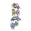
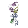
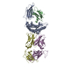
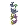
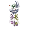
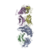
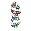
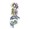
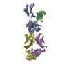
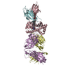
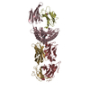
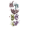
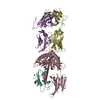
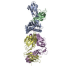
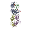
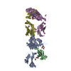
- PDB-1qrn: CRYSTAL STRUCTURE OF HUMAN A6 TCR COMPLEXED WITH HLA-A2 BOUND TO ... -
+ Open data

- Basic information
| Entry | Database: PDB / ID: 1qrn | ||||||
|---|---|---|---|---|---|---|---|
| Title | CRYSTAL STRUCTURE OF HUMAN A6 TCR COMPLEXED WITH HLA-A2 BOUND TO ALTERED HTLV-1 TAX PEPTIDE P6A | ||||||
 Components Components |
| ||||||
 Keywords Keywords |  IMMUNE SYSTEM / HUMAN TCR-PEPTIDE-MHC COMPLEX / HLA-A2 / HTLV-1 / TAX / TCR / T CELL RECEPTOR IMMUNE SYSTEM / HUMAN TCR-PEPTIDE-MHC COMPLEX / HLA-A2 / HTLV-1 / TAX / TCR / T CELL RECEPTOR | ||||||
| Function / homology |  Function and homology information Function and homology informationalpha-beta T cell receptor complex / Translocation of ZAP-70 to Immunological synapse / Phosphorylation of CD3 and TCR zeta chains / T cell mediated cytotoxicity directed against tumor cell target / antigen processing and presentation of endogenous peptide antigen via MHC class I via ER pathway, TAP-dependent / positive regulation of memory T cell activation / alpha-beta T cell activation / Generation of second messenger molecules / TAP complex binding / antigen processing and presentation of exogenous peptide antigen via MHC class I ...alpha-beta T cell receptor complex / Translocation of ZAP-70 to Immunological synapse / Phosphorylation of CD3 and TCR zeta chains / T cell mediated cytotoxicity directed against tumor cell target / antigen processing and presentation of endogenous peptide antigen via MHC class I via ER pathway, TAP-dependent / positive regulation of memory T cell activation / alpha-beta T cell activation / Generation of second messenger molecules / TAP complex binding / antigen processing and presentation of exogenous peptide antigen via MHC class I / Golgi medial cisterna / positive regulation of CD8-positive, alpha-beta T cell activation / CD8-positive, alpha-beta T cell activation / PD-1 signaling / positive regulation of CD8-positive, alpha-beta T cell proliferation / CD8 receptor binding / endoplasmic reticulum exit site / beta-2-microglobulin binding / TAP binding / protection from natural killer cell mediated cytotoxicity / antigen processing and presentation of endogenous peptide antigen via MHC class I via ER pathway, TAP-independent / antigen processing and presentation of endogenous peptide antigen via MHC class Ib / detection of bacterium / T cell receptor binding / positive regulation of ferrous iron binding / positive regulation of transferrin receptor binding / early endosome lumen / positive regulation of receptor binding / Nef mediated downregulation of MHC class I complex cell surface expression / DAP12 interactions / negative regulation of receptor binding / lumenal side of endoplasmic reticulum membrane / response to bacterium / Endosomal/Vacuolar pathway / Antigen Presentation: Folding, assembly and peptide loading of class I MHC / antigen processing and presentation of exogenous protein antigen via MHC class Ib, TAP-dependent / cellular response to iron(III) ion / negative regulation of forebrain neuron differentiation / ER to Golgi transport vesicle membrane / response to molecule of bacterial origin / regulation of erythrocyte differentiation / regulation of iron ion transport / MHC class I peptide loading complex / HFE-transferrin receptor complex / T cell mediated cytotoxicity / cellular response to iron ion / antigen processing and presentation of endogenous peptide antigen via MHC class I / positive regulation of T cell cytokine production / MHC class I protein complex / multicellular organismal-level iron ion homeostasis / positive regulation of T cell mediated cytotoxicity / peptide antigen assembly with MHC class II protein complex / negative regulation of neurogenesis / positive regulation of receptor-mediated endocytosis / MHC class II protein complex / cellular response to nicotine / recycling endosome membrane / phagocytic vesicle membrane / specific granule lumen / peptide antigen binding / positive regulation of cellular senescence / antigen processing and presentation of exogenous peptide antigen via MHC class II / Immunoregulatory interactions between a Lymphoid and a non-Lymphoid cell / Interferon gamma signaling / positive regulation of immune response / negative regulation of epithelial cell proliferation / Modulation by Mtb of host immune system / positive regulation of T cell activation / Interferon alpha/beta signaling / positive regulation of type II interferon production / sensory perception of smell / negative regulation of neuron projection development / Downstream TCR signaling / E3 ubiquitin ligases ubiquitinate target proteins / tertiary granule lumen / DAP12 signaling / MHC class II protein complex binding / T cell differentiation in thymus / late endosome membrane / positive regulation of protein binding / ER-Phagosome pathway / iron ion transport / antibacterial humoral response / T cell receptor signaling pathway / protein refolding / early endosome membrane / protein homotetramerization / intracellular iron ion homeostasis / amyloid fibril formation / adaptive immune response / learning or memory / defense response to Gram-positive bacterium / immune response / Amyloid fiber formation / lysosomal membrane / endoplasmic reticulum lumen / external side of plasma membrane / Golgi membrane / signaling receptor binding / focal adhesionSimilarity search - Function | ||||||
| Biological species |  Homo sapiens (human) Homo sapiens (human) | ||||||
| Method | X-RAY DIFFRACTION / Resolution: 2.8 Å | ||||||
 Authors Authors | Ding, Y.H. / Baker, B.M. / Garboczi, D.N. / Biddison, W.E. / Wiley, D.C. | ||||||
 Citation Citation | Journal: Immunity / Year: 1999 Title: Four A6-TCR/peptide/HLA-A2 structures that generate very different T cell signals are nearly identical. Authors: Ding, Y.H. / Baker, B.M. / Garboczi, D.N. / Biddison, W.E. / Wiley, D.C. #1: Journal: Nature / Year: 1996Title: Structure of the Complex between Human T-Cell Receptor, Viral Peptide and Hla-A2 Authors: Garboczi, D.N. / Ghosh, P. / Utz, U. / Fan, Q.R. / Wiley, D.C. | ||||||
| History |
|
- Structure visualization
| Structure viewer | Molecule: MolmilJmol/JSmol |
|---|
- Downloads & links
-Download
| PDBx/mmCIF format | 1qrn.cif.gz | 172.1 KB | Display | PDBx/mmCIF format |
|---|---|---|---|---|
| PDB format | pdb1qrn.ent.gz | 140.9 KB | Display | PDB format |
| PDBx/mmJSON format | 1qrn.json.gz | Tree view | PDBx/mmJSON format | |
| Others |  Other downloads Other downloads |
-Validation report
| Arichive directory | https://data.pdbj.org/pub/pdb/validation_reports/qr/1qrnftp://data.pdbj.org/pub/pdb/validation_reports/qr/1qrn | HTTPS FTP |
|---|
-Related structure data


-Links
 PDBj
PDBj


- Assembly
| Deposited unit | 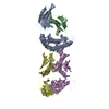
| ||||||||||
|---|---|---|---|---|---|---|---|---|---|---|---|
| 1 |
| ||||||||||
| Unit cell |
|
-Components
-Protein , 2 types, 2 molecules AB
| #1: Protein | Mass: 31725.088 Da / Num. of mol.: 1 Source method: isolated from a genetically manipulated source Source: (gene. exp.) Homo sapiens (human) / Production host:  Escherichia coli (E. coli) / References: UniProt: P01892, UniProt: P04439*PLUS Escherichia coli (E. coli) / References: UniProt: P01892, UniProt: P04439*PLUS |
|---|---|
| #2: Protein | Mass: 11879.356 Da / Num. of mol.: 1 Source method: isolated from a genetically manipulated source Source: (gene. exp.) Homo sapiens (human) / Production host: Escherichia coli (E. coli) / References: UniProt: P61769 |
-T-CELL RECEPTOR, ... , 2 types, 2 molecules DE
| #4: Protein | / A6-TCR Mass: 22130.264 Da / Num. of mol.: 1 Source method: isolated from a genetically manipulated source Source: (gene. exp.) Homo sapiens (human) / Production host: Escherichia coli (E. coli) / References: UniProt: P01848 |
|---|---|
| #5: Protein | / TCR BETA CHAIN Mass: 27175.199 Da / Num. of mol.: 1 Source method: isolated from a genetically manipulated source Source: (gene. exp.) Homo sapiens (human) / Production host: Escherichia coli (E. coli) |
-Protein/peptide / Non-polymers , 2 types, 52 molecules C
| #3: Protein/peptide | Mass: 1044.243 Da / Num. of mol.: 1 / Source method: obtained synthetically |
|---|---|
| #6: Water | ChemComp-HOH / WaterMass: 18.015 Da / Num. of mol.: 51 / Source method: isolated from a natural source / Formula: H2O |
-Experimental details
-Experiment
| Experiment | Method: X-RAY DIFFRACTION / Number of used crystals: 1 |
|---|
- Sample preparation
| Crystal | Density Matthews: 2.81 Å3/Da / Density % sol: 56.26 % | ||||||||||||||||||||||||||||||||||||
|---|---|---|---|---|---|---|---|---|---|---|---|---|---|---|---|---|---|---|---|---|---|---|---|---|---|---|---|---|---|---|---|---|---|---|---|---|---|
| Crystal grow | Temperature: 287 K / Method: vapor diffusion, hanging drop / pH: 7 Details: seeding with hanging drop vapor diffusion; 50 mM MOPS pH 7.0, 75 mM MgSO4, 13% PEG 8000, VAPOR DIFFUSION, HANGING DROP, temperature 287K | ||||||||||||||||||||||||||||||||||||
| Crystal grow | *PLUS pH: 6.5 / Method: unknown | ||||||||||||||||||||||||||||||||||||
| Components of the solutions | *PLUS
|
-Data collection
| Diffraction | Mean temperature: 110 K |
|---|---|
| Diffraction source | Source: ROTATING ANODE / Type: ELLIOTT GX-13 / Wavelength: 1.5418 |
| Detector | Type: MARRESEARCH / Detector: AREA DETECTOR / Date: Nov 26, 1997 |
| Radiation | Protocol: SINGLE WAVELENGTH / Monochromatic (M) / Laue (L): M / Scattering type: x-ray |
| Radiation wavelength | Wavelength: 1.5418 Å / Relative weight: 1 |
| Reflection | Resolution: 2.8→25 Å / Num. all: 25985 / Num. obs: 25752 / Redundancy: 3.1 % / Rmerge(I) obs: 0.076 / Net I/σ(I): 15.1 |
| Reflection shell | Resolution: 2.8→2.9 Å / Redundancy: 2.5 % / Rmerge(I) obs: 0.26 |
| Reflection | *PLUS Lowest resolution: 25 Å / % possible obs: 98.2 % |
| Reflection shell | *PLUS % possible obs: 83.2 % / Mean I/σ(I) obs: 3.7 |
- Processing
| Software |
| ||||||||||||||||||||||||||||||
|---|---|---|---|---|---|---|---|---|---|---|---|---|---|---|---|---|---|---|---|---|---|---|---|---|---|---|---|---|---|---|---|
| Refinement | Resolution: 2.8→25 Å / Cross valid method: THROUGHOUT / σ(F): 0 / σ(I): 0
| ||||||||||||||||||||||||||||||
| Refinement step | Cycle: LAST / Resolution: 2.8→25 Å
| ||||||||||||||||||||||||||||||
| Software | *PLUS Name: CNS / Classification: refinement | ||||||||||||||||||||||||||||||
| Refinement | *PLUS Highest resolution: 2.8 Å / Lowest resolution: 25 Å / σ(F): 0 / % reflection Rfree: 10.5 % / Rfactor obs: 0.216 | ||||||||||||||||||||||||||||||
| Solvent computation | *PLUS | ||||||||||||||||||||||||||||||
| Displacement parameters | *PLUS | ||||||||||||||||||||||||||||||
| Refine LS restraints | *PLUS
|
