- PDB-1qle: CRYO-STRUCTURE OF THE PARACOCCUS DENITRIFICANS FOUR-SUBUNIT CYTOC... -
+
Open data
ID or keywords:
Loading...
-
Basic information
Entry
Database: PDB / ID: 1qle
Title
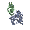
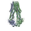
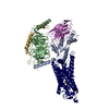
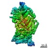
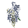
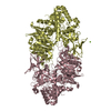
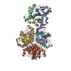
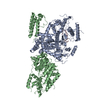
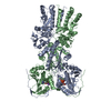
CRYO-STRUCTURE OF THE PARACOCCUS DENITRIFICANS FOUR-SUBUNIT CYTOCHROME C OXIDASE IN THE COMPLETELY OXIDIZED STATE COMPLEXED WITH AN ANTIBODY FV FRAGMENT
Components
(CYTOCHROME C OXIDASE POLYPEPTIDE ...) x 3
CCYTOCHROME C OXIDASE
HEAVY CHAIN ANTIBODY FV FRAGMENT
LIGHT CHAIN ANTIBODY FV FRAGMENT
Keywords
OXIDOREDUCTASE/IMMUNE SYSTEM / COMPLEX (OXIDOREDUCTASE-ANTIBODY) / ELECTRON TRANSPORT / TRANSMEMBRANE / CYTOCHROME OXIDASE / ANTIBODY COMPLEX / OXIDOREDUCTASE-IMMUNE SYSTEM complex
Function / homology
Function and homology information
respiratory chain complex IV / aerobic electron transport chain / cytochrome-c oxidase / oxidative phosphorylation / electron transport coupled proton transport / cytochrome-c oxidase activity / respirasome / copper ion binding / heme binding / metal ion binding / plasma membrane Similarity search - Function
Bacterial aa3 type cytochrome c oxidase subunit IV / Cytochrome c oxidase, subunit IV, bacterial aa3 type / Bacterial aa3 type cytochrome c oxidase subunit IV superfamily / Bacterial aa3 type cytochrome c oxidase subunit IV / Cytochrome c oxidase, subunit I bacterial type / Cytochrome c oxidase, subunit III, four-helix bundle / Helix Hairpins - #90 / Cytochrome C Oxidase; Chain A / Cytochrome c oxidase-like, subunit I domain / Cytochrome c oxidase subunit III domain ...Bacterial aa3 type cytochrome c oxidase subunit IV / Cytochrome c oxidase, subunit IV, bacterial aa3 type / Bacterial aa3 type cytochrome c oxidase subunit IV superfamily / Bacterial aa3 type cytochrome c oxidase subunit IV / Cytochrome c oxidase, subunit I bacterial type / Cytochrome c oxidase, subunit III, four-helix bundle / Helix Hairpins - #90 / Cytochrome C Oxidase; Chain A / Cytochrome c oxidase-like, subunit I domain / Cytochrome c oxidase subunit III domain / Cytochrome c oxidase subunit I domain / Cytochrome c oxidase, subunit II / Cytochrome C oxidase subunit II, transmembrane domain / Cytochrome c oxidase subunit III / Cytochrome c oxidase subunit III-like / Cytochrome c oxidase, subunit III, 4-helical bundle / Cytochrome c oxidase subunit III / Heme-copper oxidase subunit III family profile. / Cytochrome c oxidase subunit III-like superfamily / Cytochrome C oxidase subunit II, transmembrane domain / Cytochrome oxidase subunit II transmembrane region profile. / Cytochrome c/quinol oxidase subunit II / Copper centre Cu(A) / Cytochrome C oxidase subunit II, transmembrane domain superfamily / CO II and nitrous oxide reductase dinuclear copper centers signature. / Cytochrome c oxidase, subunit I, copper-binding site / Heme-copper oxidase catalytic subunit, copper B binding region signature. / Cytochrome c oxidase-like, subunit I domain / Cytochrome oxidase subunit I profile. / Cytochrome c oxidase subunit I / Cytochrome c oxidase-like, subunit I superfamily / Cytochrome C and Quinol oxidase polypeptide I / Cytochrome C oxidase subunit II, periplasmic domain / Cytochrome c oxidase subunit II-like C-terminal / Cytochrome oxidase subunit II copper A binding domain profile. / Helix Hairpins - #70 / Cupredoxins - blue copper proteins / Four Helix Bundle (Hemerythrin (Met), subunit A) / Cupredoxin / Single alpha-helices involved in coiled-coils or other helix-helix interfaces / Helix Hairpins / Immunoglobulins / Up-down Bundle / Immunoglobulin-like / Sandwich / Orthogonal Bundle / Mainly Beta / Mainly Alpha Similarity search - Domain/homology
COPPER (II) ION / DINUCLEAR COPPER ION / HEME-A / : / 1,2-DIACYL-SN-GLYCERO-3-PHOSPHOCHOLINE / Cytochrome c oxidase subunit 3 / Cytochrome c oxidase subunit 2 / Cytochrome c oxidase subunit 4 / Cytochrome c oxidase subunit 1-beta Similarity search - Component
A: CYTOCHROME C OXIDASE POLYPEPTIDE I-BETA B: CYTOCHROME C OXIDASE POLYPEPTIDE II C: CYTOCHROME C OXIDASE POLYPEPTIDE III D: CCYTOCHROME C OXIDASE H: HEAVY CHAIN ANTIBODY FV FRAGMENT L: LIGHT CHAIN ANTIBODY FV FRAGMENT hetero molecules
THE CRYSTAL STRUCTURE SHOWS A CRYSTAL PACKING ARRANGEMENT WHERETHERE IS CONTACT BETWEEN CHAINS C AND H GIVING A CYCLICPACKING WITHCHAIN- H ... (CHAIN-C...CHAIN-H) ... CHAIN-C(Y,-X,Z ) ASU: X,Y,Z (-Y,X,Z)
Mass: 30676.975 Da / Num. of mol.: 1 / Source method: isolated from a natural source / Source: (natural) PARACOCCUS DENITRIFICANS (bacteria) / Cellular location: CYTOPLASMIC MEMBRANECell membrane / References: UniProt: P06030, cytochrome-c oxidase
-
Protein/peptide , 1 types, 1 molecules D
#4: Protein/peptide
CCYTOCHROMECOXIDASE / CYTOCHROME AA3
Mass: 4701.407 Da / Num. of mol.: 1 / Source method: isolated from a natural source / Source: (natural) PARACOCCUS DENITRIFICANS (bacteria) / Cellular location: CYTOPLASMIC MEMBRANECell membrane / References: UniProt: P77921, cytochrome-c oxidase
-
Antibody , 2 types, 2 molecules HL
#5: Antibody
HEAVYCHAINANTIBODYFVFRAGMENT
Mass: 13356.854 Da / Num. of mol.: 1 Source method: isolated from a genetically manipulated source Source: (gene. exp.) MUS MUSCULUS (house mouse) / Production host: ESCHERICHIA COLI (E. coli)
#6: Antibody
LIGHTCHAINANTIBODYFVFRAGMENT
Mass: 11786.118 Da / Num. of mol.: 1 Source method: isolated from a genetically manipulated source Source: (gene. exp.) MUS MUSCULUS (house mouse) / Production host: ESCHERICHIA COLI (E. coli)
Mass: 790.145 Da / Num. of mol.: 2 / Source method: obtained synthetically / Formula: C44H88NO8P / Comment: phospholipid*YM
-
Details
Sequence details
THE SEQUENCE OF VH AND VL IS DESCRIBED IN THE FOLLOWING REFERENCE, OSTERMEIER C. ET AL. (1995) ...THE SEQUENCE OF VH AND VL IS DESCRIBED IN THE FOLLOWING REFERENCE, OSTERMEIER C. ET AL. (1995) PROTEINS 21: 74-77. THE NCBI REFERENCE LOCUS FOR CHAIN H IS 2914143 THE NCBI REFERENCE LOCUS FOR CHAIN L IS 2914144
-
Experimental details
-
Experiment
Experiment
Method: X-RAY DIFFRACTION / Number of used crystals: 2
-
Sample preparation
Crystal
Density Matthews: 5.74 Å3/Da / Density % sol: 75 %
Crystal grow
pH: 8 / Details: pH 8.00
Crystal grow
*PLUS
Temperature: 14 ℃ / Method: vapor diffusion, sitting drop
Protocol: SINGLE WAVELENGTH / Monochromatic (M) / Laue (L): M / Scattering type: x-ray
Radiation wavelength
Wavelength: 0.9875 Å / Relative weight: 1
Reflection
Resolution: 3→20 Å / Num. obs: 64653 / % possible obs: 95.2 % / Observed criterion σ(I): 0 / Redundancy: 2.5 % / Biso Wilson estimate: 32.2 Å2 / Rmerge(I) obs: 0.115 / Net I/σ(I): 9.6
Reflection
*PLUS
Num. measured all: 165650
-
Processing
Software
Name
Version
Classification
CNS
0.3
refinement
DENZO
datareduction
SCALEPACK
datascaling
CNS
0.3
phasing
Refinement
Method to determine structure: OTHER / Resolution: 3→6 Å / Rfactor Rfree error: 0.007 / Data cutoff high absF: 5840810 / Cross valid method: THROUGHOUT / σ(F): 2
Rfactor
Num. reflection
% reflection
Selection details
Rfree
0.309
1975
5 %
RANDOM
Rwork
0.235
-
-
-
obs
0.235
39276
66.2 %
-
Displacement parameters
Biso mean: 31.5 Å2
Baniso -1
Baniso -2
Baniso -3
1-
-0.39 Å2
0 Å2
0 Å2
2-
-
-0.39 Å2
0 Å2
3-
-
-
0.78 Å2
Refine analyze
Free
Obs
Luzzati coordinate error
0.55 Å
0.4 Å
Luzzati d res low
-
5 Å
Luzzati sigma a
0.8 Å
0.44 Å
Refinement step
Cycle: LAST / Resolution: 3→6 Å
Protein
Nucleic acid
Ligand
Solvent
Total
Num. atoms
10529
0
233
0
10762
Refine LS restraints
Refine-ID
Type
Dev ideal
X-RAY DIFFRACTION
c_bond_d
0.012
X-RAY DIFFRACTION
c_bond_d_na
X-RAY DIFFRACTION
c_bond_d_prot
X-RAY DIFFRACTION
c_angle_d
X-RAY DIFFRACTION
c_angle_d_na
X-RAY DIFFRACTION
c_angle_d_prot
X-RAY DIFFRACTION
c_angle_deg
1.6
X-RAY DIFFRACTION
c_angle_deg_na
X-RAY DIFFRACTION
c_angle_deg_prot
X-RAY DIFFRACTION
c_dihedral_angle_d
23.8
X-RAY DIFFRACTION
c_dihedral_angle_d_na
X-RAY DIFFRACTION
c_dihedral_angle_d_prot
X-RAY DIFFRACTION
c_improper_angle_d
1.5
X-RAY DIFFRACTION
c_improper_angle_d_na
X-RAY DIFFRACTION
c_improper_angle_d_prot
X-RAY DIFFRACTION
c_mcbond_it
X-RAY DIFFRACTION
c_mcangle_it
X-RAY DIFFRACTION
c_scbond_it
X-RAY DIFFRACTION
c_scangle_it
LS refinement shell
Resolution: 3→3.16 Å / Rfactor Rfree error: 0.026 / Total num. of bins used: 6
In the structure databanks used in Yorodumi, some data are registered as the other names, "COVID-19 virus" and "2019-nCoV". Here are the details of the virus and the list of structure data.
Jan 31, 2019. EMDB accession codes are about to change! (news from PDBe EMDB page)
EMDB accession codes are about to change! (news from PDBe EMDB page)
The allocation of 4 digits for EMDB accession codes will soon come to an end. Whilst these codes will remain in use, new EMDB accession codes will include an additional digit and will expand incrementally as the available range of codes is exhausted. The current 4-digit format prefixed with “EMD-” (i.e. EMD-XXXX) will advance to a 5-digit format (i.e. EMD-XXXXX), and so on. It is currently estimated that the 4-digit codes will be depleted around Spring 2019, at which point the 5-digit format will come into force.
The EM Navigator/Yorodumi systems omit the EMD- prefix.
Related info.:Q: What is EMD? / ID/Accession-code notation in Yorodumi/EM Navigator
Yorodumi is a browser for structure data from EMDB, PDB, SASBDB, etc.
This page is also the successor to EM Navigator detail page, and also detail information page/front-end page for Omokage search.
The word "yorodu" (or yorozu) is an old Japanese word meaning "ten thousand". "mi" (miru) is to see.
Related info.:EMDB / PDB / SASBDB / Comparison of 3 databanks / Yorodumi Search / Aug 31, 2016. New EM Navigator & Yorodumi / Yorodumi Papers / Jmol/JSmol / Function and homology information / Changes in new EM Navigator and Yorodumi
 Movie
Movie Controller

 Yorodumi
 Open data
 Basic information
 Components
 Keywords
 ELECTRON TRANSPORT /
  Function and homology information

 Authors
 Citation
 Structure visualization
 Downloads & links
 Other downloads

 PDBj
PDBj


 Assembly


 Mass: 852.837 Da / Num. of mol.: 2 / Source method: obtained synthetically / Formula: C49H56FeN4O6
 Mass: 63.546 Da / Num. of mol.: 1 / Source method: obtained synthetically / Formula: Cu
 Mass: 40.078 Da / Num. of mol.: 1 / Source method: obtained synthetically / Formula: Ca
 Mass: 54.938 Da / Num. of mol.: 1 / Source method: obtained synthetically / Formula: Mn
 Mass: 127.092 Da / Num. of mol.: 1 / Source method: obtained synthetically / Formula: Cu2
 Mass: 790.145 Da / Num. of mol.: 2 / Source method: obtained synthetically / Formula: C44H88NO8P / Comment: phospholipid*YM
 Sample preparation
 / Beamline: ID2 / Wavelength: 0.9875
  Processing