 Movie
Movie Controller

[English] 日本語
 Yorodumi
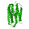
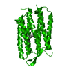
- PDB-1qkp: HIGH RESOLUTION X-RAY STRUCTURE OF AN EARLY INTERMEDIATE IN THE B... -
+ Open data

- Basic information
| Entry | Database: PDB / ID: 1qkp | |||||||||
|---|---|---|---|---|---|---|---|---|---|---|
| Title | HIGH RESOLUTION X-RAY STRUCTURE OF AN EARLY INTERMEDIATE IN THE BACTERIORHODOPSIN PHOTOCYCLE | |||||||||
 Components Components | BACTERIORHODOPSIN | |||||||||
 Keywords Keywords | PHOTORECEPTOR / PROTON PUMP / MEMBRANE PROTEIN / RETINAL PROTEIN / INTERMEDIATE STATE / PHOTOCYCLE / LIPIDIC CUBIC PHASES | |||||||||
| Function / homology |  Function and homology informationphotoreceptor activity / phototransduction / proton transmembrane transport / monoatomic ion channel activity / plasma membrane Function and homology informationphotoreceptor activity / phototransduction / proton transmembrane transport / monoatomic ion channel activity / plasma membraneSimilarity search - Function | |||||||||
| Biological species |  HALOBACTERIUM SALINARIUM (Halophile) HALOBACTERIUM SALINARIUM (Halophile) | |||||||||
| Method | X-RAY DIFFRACTION / SYNCHROTRON / MOLECULAR REPLACEMENT / Resolution: 2.1 Å | |||||||||
 Authors Authors | Edman, K. / Nollert, P. / Royant, A. / Belrhali, H. / Pebay-Peyroula, E. / Hajdu, J. / Neutze, R. / Landau, E.M. | |||||||||
 Citation Citation | Journal: Nature / Year: 1999 Title: High-resolution X-ray structure of an early intermediate in the bacteriorhodopsin photocycle. Authors: Edman, K. / Nollert, P. / Royant, A. / Belrhali, H. / Pebay-Peyroula, E. / Hajdu, J. / Neutze, R. / Landau, E.M. #1: Journal: Structure / Year: 1999Title: Protein, Lipid and Water Organization in Bacteriorhodopsin: A Molecular View of the Purple Membrane at 1.9 Angstrom Resolution Authors: Belrhali, H. / Nollert, P. / Royant, A. / Menzel, C. / Rosenbusch, J.P. / Landau, E.M. / Pebay-Peyroula, E. | |||||||||
| History |
| |||||||||
| Remark 650 | HELIX DETERMINATION METHOD: DSSP | |||||||||
| Remark 700 | SHEET DETERMINATION METHOD: DSSP |
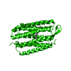
- Structure visualization
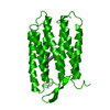
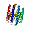
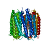
| Structure viewer | Molecule: MolmilJmol/JSmol |
|---|
- Downloads & links
-Download
| PDBx/mmCIF format | 1qkp.cif.gz | 60.3 KB | Display | PDBx/mmCIF format |
|---|---|---|---|---|
| PDB format | pdb1qkp.ent.gz | 44.1 KB | Display | PDB format |
| PDBx/mmJSON format | 1qkp.json.gz | Tree view | PDBx/mmJSON format | |
| Others |  Other downloads Other downloads |
-Validation report
| Arichive directory | https://data.pdbj.org/pub/pdb/validation_reports/qk/1qkpftp://data.pdbj.org/pub/pdb/validation_reports/qk/1qkp | HTTPS FTP |
|---|
-Related structure data
| Related structure data |  1qkoC  1qhjS C: citing same article ( S: Starting model for refinement |
|---|---|
| Similar structure data |


-Links
 PDBj
PDBj


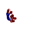
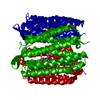
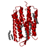
- Assembly
| Deposited unit | 
| ||||||||
|---|---|---|---|---|---|---|---|---|---|
| 1 | 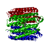
| ||||||||
| Unit cell |
| ||||||||
| Details | A XRAY OBSERVED TRIMERIC BIO- ASSEMBLY CAN BE BUILTBY SPACE GROUP SYMMETRY EXPANSION OF THECONTENTS OF THE ASYMMETRIC UNIT |
-Components
| #1: Protein | Mass: 26814.412 Da / Num. of mol.: 1 / Source method: isolated from a natural source / Details: RETINAL LINKED TO LYS 216 VIA A SCHIFF BASE / Source: (natural) HALOBACTERIUM SALINARIUM (Halophile) / Cellular location: PLASMA MEMBRANECell membrane / Strain: S9 / References: UniProt: P02945 |
|---|---|
| #2: Chemical | ChemComp-RET / Retinal  Mass: 284.436 Da / Num. of mol.: 1 / Source method: obtained synthetically / Formula: C20H28O Mass: 284.436 Da / Num. of mol.: 1 / Source method: obtained synthetically / Formula: C20H28O |
| #3: Water | ChemComp-HOH / Water Mass: 18.015 Da / Num. of mol.: 27 / Source method: isolated from a natural source / Formula: H2O Mass: 18.015 Da / Num. of mol.: 27 / Source method: isolated from a natural source / Formula: H2O |
-Experimental details
-Experiment
| Experiment | Method: X-RAY DIFFRACTION / Number of used crystals: 3 |
|---|
- Sample preparation
| Crystal | Density Matthews: 2.39 Å3/Da / Density % sol: 48 % Description: THE CRYSTAL FORM WAS THE SAME AS FOR 1QHJ, THEREFORE 1QHJ WAS DIRECTLY USED FOR THE INITIAL PHASING | |||||||||||||||||||||||||
|---|---|---|---|---|---|---|---|---|---|---|---|---|---|---|---|---|---|---|---|---|---|---|---|---|---|---|
| Crystal grow | Temperature: 293 K / Method: lipidic cubic phase / pH: 5.6 Details: PROTEIN FROM THE PURPLE MEMBRANE WAS DELIPIDATED AND SOLUBILIZED IN OCTYL GLUCOSIDE. PROTEIN WAS CRYSTALLIZED FROM 60 - 70% (W/W) MONOOLEIN, 0.7 - 4.0 M NA/K - PHOSPHATE IN A PHOSPHATE ...Details: PROTEIN FROM THE PURPLE MEMBRANE WAS DELIPIDATED AND SOLUBILIZED IN OCTYL GLUCOSIDE. PROTEIN WAS CRYSTALLIZED FROM 60 - 70% (W/W) MONOOLEIN, 0.7 - 4.0 M NA/K - PHOSPHATE IN A PHOSPHATE BUFFER AT PH 5.6, AT 20C AND IN THE DARK. THE MIXTURE WAS CENTRIFUGED AT 10000G FOR 150 MN PRIOR TO CRYSTALLISATION. | |||||||||||||||||||||||||
| Crystal | *PLUS | |||||||||||||||||||||||||
| Crystal grow | *PLUS Temperature: 20 ℃ / Method: unknownDetails: Landau, E.M., (1996) Proc.Natl.Acad.Sci.USA., 93, 14532. | |||||||||||||||||||||||||
| Components of the solutions | *PLUS
|
-Data collection
| Diffraction | Mean temperature: 110 K |
|---|---|
| Diffraction source | Source: SYNCHROTRON / Site: ESRF  / Beamline: ID14-3 / Wavelength: 0.936 / Beamline: ID14-3 / Wavelength: 0.936 |
| Detector | Type: MARRESEARCH / Detector: CCD / Date: Feb 15, 1999 |
| Radiation | Protocol: SINGLE WAVELENGTH / Monochromatic (M) / Laue (L): M / Scattering type: x-ray |
| Radiation wavelength | Wavelength: 0.936 Å / Relative weight: 1 |
| Reflection | Resolution: 2.1→38 Å / Num. obs: 13653 / % possible obs: 99.8 % / Observed criterion σ(I): 0 / Redundancy: 4.9 % / Biso Wilson estimate: 35 Å2 / Rsym value: 0.055 / Net I/σ(I): 10.1 |
| Reflection shell | Resolution: 2.1→2.2 Å / Redundancy: 4.9 % / Mean I/σ(I) obs: 2.1 / Rsym value: 0.37 / % possible all: 99.6 |
| Reflection | *PLUS Num. measured all: 67437 / Rmerge(I) obs: 0.055 |
| Reflection shell | *PLUS % possible obs: 99.6 % / Rmerge(I) obs: 0.37 |
- Processing
| Software |
| ||||||||||||||||||||||||||||||||||||||||||||||||||||||||||||||||||||||||||||||||
|---|---|---|---|---|---|---|---|---|---|---|---|---|---|---|---|---|---|---|---|---|---|---|---|---|---|---|---|---|---|---|---|---|---|---|---|---|---|---|---|---|---|---|---|---|---|---|---|---|---|---|---|---|---|---|---|---|---|---|---|---|---|---|---|---|---|---|---|---|---|---|---|---|---|---|---|---|---|---|---|---|---|
| Refinement | Method to determine structure: MOLECULAR REPLACEMENT Starting model: 1QHJ Resolution: 2.1→38 Å / Data cutoff high absF: 10000 / Cross valid method: THROUGHOUT / σ(F): 0 / Stereochemistry target values: MLF Details: THIS STRUCTURE WAS REFINED FROM AN ILLUMINATED CRYSTAL AND REPRESENTS A MIXTURE OF THE GROUND STATE AND THE LOW TEMPERATURE K STATE OF BACTERIORHODOPSIN. DURING THE DATA COLLECTION, THE ...Details: THIS STRUCTURE WAS REFINED FROM AN ILLUMINATED CRYSTAL AND REPRESENTS A MIXTURE OF THE GROUND STATE AND THE LOW TEMPERATURE K STATE OF BACTERIORHODOPSIN. DURING THE DATA COLLECTION, THE CRYSTAL WAS MAINTAINED AT 110 K AND CONTINUOUSLY ILLUMINATED WITH A DIODE LASER AT 532 NM. THREE DATA SETS FROM ILLUMINATED CRYSTALS WERE COLLECTED. FROM THE 3 EXPERIMENTAL DIFFERENCE MAPS , FEXC-FGROUND (DATA IN R1QHJSF), 9 RESIDUES SHOWED SIGNIFICANT CHANGES IN THE EXCITED STATE. A FIRST REFINEMENT ON THE REMAINING RESIDUES WAS PERFORMED, FIXING THE 9 RESIDUES. THEN THESE 9 RESIDUES WERE REFINED WITH ALTERNATE CONFORMATIONS FIXING THE REMAINING STRUCTURE.
| ||||||||||||||||||||||||||||||||||||||||||||||||||||||||||||||||||||||||||||||||
| Solvent computation | Solvent model: FLAT MODEL / Bsol: 90 Å2 / ksol: 0.399 e/Å3 | ||||||||||||||||||||||||||||||||||||||||||||||||||||||||||||||||||||||||||||||||
| Displacement parameters | Biso mean: 36.5 Å2
| ||||||||||||||||||||||||||||||||||||||||||||||||||||||||||||||||||||||||||||||||
| Refine analyze |
| ||||||||||||||||||||||||||||||||||||||||||||||||||||||||||||||||||||||||||||||||
| Refinement step | Cycle: LAST / Resolution: 2.1→38 Å
| ||||||||||||||||||||||||||||||||||||||||||||||||||||||||||||||||||||||||||||||||
| Refine LS restraints |
| ||||||||||||||||||||||||||||||||||||||||||||||||||||||||||||||||||||||||||||||||
| LS refinement shell | Resolution: 2.1→2.18 Å / Total num. of bins used: 10
| ||||||||||||||||||||||||||||||||||||||||||||||||||||||||||||||||||||||||||||||||
| Xplor file |
| ||||||||||||||||||||||||||||||||||||||||||||||||||||||||||||||||||||||||||||||||
| Software | *PLUS Name: CNS / Version: 0.5 / Classification: refinement | ||||||||||||||||||||||||||||||||||||||||||||||||||||||||||||||||||||||||||||||||
| Refinement | *PLUS Rfactor Rfree: 0.255 | ||||||||||||||||||||||||||||||||||||||||||||||||||||||||||||||||||||||||||||||||
| Solvent computation | *PLUS | ||||||||||||||||||||||||||||||||||||||||||||||||||||||||||||||||||||||||||||||||
| Displacement parameters | *PLUS | ||||||||||||||||||||||||||||||||||||||||||||||||||||||||||||||||||||||||||||||||
| Refine LS restraints | *PLUS
|
