 Movie
Movie Controller

[English] 日本語
 Yorodumi
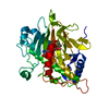
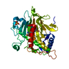
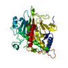
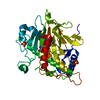
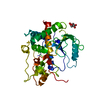
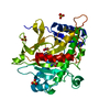
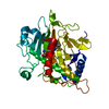
- PDB-1qjf: ISOPENICILLIN N SYNTHASE FROM ASPERGILLUS NIDULANS (Monocyclic Su... -
+ Open data

- Basic information
| Entry | Database: PDB / ID: 1qjf | ||||||
|---|---|---|---|---|---|---|---|
| Title | ISOPENICILLIN N SYNTHASE FROM ASPERGILLUS NIDULANS (Monocyclic Sulfoxide - Fe COMPLEX) | ||||||
 Components Components | ISOPENICILLIN N SYNTHASE | ||||||
 Keywords Keywords | B-LACTAM ANTIBIOTIC / MONOCYCLIC INTERMEDIATE / OXYGENASE / PENICILLIN BIOSYNTHESIS / SULFOXIDE | ||||||
| Function / homology |  Function and homology informationisopenicillin-N synthase / isopenicillin-N synthase activity / penicillin biosynthetic process / L-ascorbic acid binding / iron ion binding / cytosol Function and homology informationisopenicillin-N synthase / isopenicillin-N synthase activity / penicillin biosynthetic process / L-ascorbic acid binding / iron ion binding / cytosolSimilarity search - Function | ||||||
| Biological species |  EMERICELLA NIDULANS (mold) EMERICELLA NIDULANS (mold) | ||||||
| Method | X-RAY DIFFRACTION / SYNCHROTRON / MOLECULAR REPLACEMENT / Resolution: 1.4 Å | ||||||
 Authors Authors | Rutledge, P.J. / Clifton, I.J. / Burzlaff, N.I. / Roach, P.L. / Adlington, R.M. / Baldwin, J.E. | ||||||
 Citation Citation | Journal: Nature / Year: 1999 Title: The Reaction Cycle of Isopenicillin N Synthase Observed by X-Ray Diffraction Authors: Burzlaff, N.I. / Rutledge, P.J. / Clifton, I.J. / Hensgens, C.M.H. / Pickford, M. / Adlington, R.M. / Roach, P.L. / Baldwin, J.E. #1: Journal: Nature / Year: 1997Title: Structure of Isopenicillin N Synthase Complexed with Substrate and the Mechanism of Penicillin Formation Authors: Roach, P.L. / Clifton, I.J. / Hensgens, C.M. / Shibata, N. / Schofield, C.J. / Hajdu, J. / Baldwin, J.E. #2: Journal: Nature / Year: 1995Title: Crystal Structure of Isopenicillin N Synthase is the First from a New Structural Family of Enzymes Authors: Roach, P.L. / Clifton, I.J. / Fulop, V. / Harlos, K. / Barton, G.J. / Hajdu, J. / Andersson, I. / Schofield, C.J. / Baldwin, J.E. #3: Journal: Protein Sci. / Year: 1995 Title: Crystallization and Preliminary X-Ray Diffraction Studies on Recombinant Isopenicillin N Synthase from Aspergillus Nidulans Authors: Roach, P.L. / Schofield, C.J. / Baldwin, J.E. / Clifton, I.J. / Hajdu, J. | ||||||
| History |
|
- Structure visualization
| Structure viewer | Molecule: MolmilJmol/JSmol |
|---|
- Downloads & links
-Download
| PDBx/mmCIF format | 1qjf.cif.gz | 91.8 KB | Display | PDBx/mmCIF format |
|---|---|---|---|---|
| PDB format | pdb1qjf.ent.gz | 67 KB | Display | PDB format |
| PDBx/mmJSON format | 1qjf.json.gz | Tree view | PDBx/mmJSON format | |
| Others |  Other downloads Other downloads |
-Validation report
| Arichive directory | https://data.pdbj.org/pub/pdb/validation_reports/qj/1qjfftp://data.pdbj.org/pub/pdb/validation_reports/qj/1qjf | HTTPS FTP |
|---|
-Related structure data
| Related structure data |  1qiqC  1qjeC  1bk0S S: Starting model for refinement C: citing same article ( |
|---|---|
| Similar structure data |


-Links
 PDBj
PDBj


- Assembly
| Deposited unit | 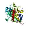
| ||||||||
|---|---|---|---|---|---|---|---|---|---|
| 1 |
| ||||||||
| Unit cell |
| ||||||||
| Details | BIOLOGICAL_UNIT: MONOMER |
-Components
| #1: Protein | Mass: 37563.836 Da / Num. of mol.: 1 Source method: isolated from a genetically manipulated source Source: (gene. exp.) EMERICELLA NIDULANS (mold) / Gene: PCB C / Plasmid: PJB703 / Production host:  ESCHERICHIA COLI (E. coli) / Strain (production host): NM554 / References: UniProt: P05326 ESCHERICHIA COLI (E. coli) / Strain (production host): NM554 / References: UniProt: P05326 |
|---|---|
| #2: Chemical | ChemComp-SO4 / Sulfate  Mass: 96.063 Da / Num. of mol.: 1 / Source method: obtained synthetically / Formula: SO4 Mass: 96.063 Da / Num. of mol.: 1 / Source method: obtained synthetically / Formula: SO4 |
| #3: Chemical | ChemComp-ACS /   Mass: 395.452 Da / Num. of mol.: 1 / Source method: obtained synthetically / Formula: C13H21N3O7S2 Mass: 395.452 Da / Num. of mol.: 1 / Source method: obtained synthetically / Formula: C13H21N3O7S2 |
| #4: Chemical | ChemComp-FE2 /   Mass: 55.845 Da / Num. of mol.: 1 / Source method: obtained synthetically / Formula: Fe Mass: 55.845 Da / Num. of mol.: 1 / Source method: obtained synthetically / Formula: Fe |
| #5: Water | ChemComp-HOH / Water Mass: 18.015 Da / Num. of mol.: 410 / Source method: isolated from a natural source / Formula: H2O Mass: 18.015 Da / Num. of mol.: 410 / Source method: isolated from a natural source / Formula: H2O |
-Experimental details
-Experiment
| Experiment | Method: X-RAY DIFFRACTION / Number of used crystals: 1 |
|---|
- Sample preparation
| Crystal | Density Matthews: 2.226 Å3/Da / Density % sol: 38 % | |||||||||||||||
|---|---|---|---|---|---|---|---|---|---|---|---|---|---|---|---|---|
| Crystal grow | pH: 8.5 Details: 1.8M LITHIUM SULPHATE, 100MM TRIS/HCL (PH8.5) (5MM FERROUS SULPHATE, 70 MM ACMC, 50MG/ML IPNS), pH 8.50 | |||||||||||||||
| Crystal grow | *PLUS Temperature: 17 ℃ / Method: vapor diffusion, hanging drop | |||||||||||||||
| Components of the solutions | *PLUS
|
-Data collection
| Diffraction | Mean temperature: 100 K |
|---|---|
| Diffraction source | Source: SYNCHROTRON / Site: EMBL/DESY, HAMBURG  / Beamline: BW7B / Wavelength: 0.8345 / Beamline: BW7B / Wavelength: 0.8345 |
| Detector | Type: MARRESEARCH / Detector: IMAGE PLATE / Date: Oct 15, 1998 |
| Radiation | Protocol: SINGLE WAVELENGTH / Monochromatic (M) / Laue (L): M / Scattering type: x-ray |
| Radiation wavelength | Wavelength: 0.8345 Å / Relative weight: 1 |
| Reflection | Resolution: 1.4→30 Å / Num. obs: 55209 / % possible obs: 85.3 % / Redundancy: 3.2 % / Rmerge(I) obs: 0.114 / Net I/σ(I): 3.6 |
| Reflection shell | Resolution: 1.4→1.48 Å / Redundancy: 1.6 % / Rmerge(I) obs: 0.343 / Mean I/σ(I) obs: 2.2 / % possible all: 50.6 |
| Reflection | *PLUS Num. measured all: 344241 |
| Reflection shell | *PLUS % possible obs: 50.6 % / Num. unique obs: 4482 / Num. measured obs: 6601 |
- Processing
| Software |
| |||||||||||||||||||||||||||||||||
|---|---|---|---|---|---|---|---|---|---|---|---|---|---|---|---|---|---|---|---|---|---|---|---|---|---|---|---|---|---|---|---|---|---|---|
| Refinement | Method to determine structure: MOLECULAR REPLACEMENT Starting model: 1BK0 Resolution: 1.4→8 Å / Num. restraintsaints: 33425 / Cross valid method: THROUGHOUT / σ(F): 0 / Details: FINAL MAP GENERATED WITH CCP4
| |||||||||||||||||||||||||||||||||
| Refine analyze | Num. disordered residues: 1 / Occupancy sum hydrogen: 2541 / Occupancy sum non hydrogen: 3059 | |||||||||||||||||||||||||||||||||
| Refinement step | Cycle: LAST / Resolution: 1.4→8 Å
| |||||||||||||||||||||||||||||||||
| Refine LS restraints |
|
