 Movie
Movie Controller

[English] 日本語
 Yorodumi
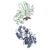
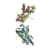
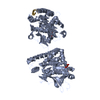
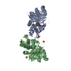
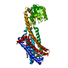
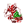
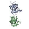
- PDB-1qh3: HUMAN GLYOXALASE II WITH CACODYLATE AND ACETATE IONS PRESENT IN T... -
+ Open data

- Basic information
| Entry | Database: PDB / ID: 1qh3 | ||||||
|---|---|---|---|---|---|---|---|
| Title | HUMAN GLYOXALASE II WITH CACODYLATE AND ACETATE IONS PRESENT IN THE ACTIVE SITE | ||||||
 Components Components | PROTEIN (HYDROXYACYLGLUTATHIONE HYDROLASE) | ||||||
 Keywords Keywords |  HYDROLASE / METALLO-HYDROLASE HYDROLASE / METALLO-HYDROLASE | ||||||
| Function / homology |  Function and homology informationhydroxyacylglutathione hydrolase / hydroxyacylglutathione hydrolase activity / methylglyoxal catabolic process to D-lactate via S-lactoyl-glutathione / glutathione biosynthetic process / Pyruvate metabolism / glutathione metabolic process / mitochondrial matrix / mitochondrion / metal ion binding / cytosol / cytoplasm Function and homology informationhydroxyacylglutathione hydrolase / hydroxyacylglutathione hydrolase activity / methylglyoxal catabolic process to D-lactate via S-lactoyl-glutathione / glutathione biosynthetic process / Pyruvate metabolism / glutathione metabolic process / mitochondrial matrix / mitochondrion / metal ion binding / cytosol / cytoplasmSimilarity search - Function | ||||||
| Biological species |  Homo sapiens (human) Homo sapiens (human) | ||||||
| Method | X-RAY DIFFRACTION / SYNCHROTRON / SIRAS / Resolution: 1.9 Å | ||||||
 Authors Authors | Cameron, A.D. / Ridderstrom, M. / Olin, B. / Mannervik, B. | ||||||
 Citation Citation | Journal: Structure Fold.Des. / Year: 1999 Title: Crystal structure of human glyoxalase II and its complex with a glutathione thiolester substrate analogue. Authors: Cameron, A.D. / Ridderstrom, M. / Olin, B. / Mannervik, B. | ||||||
| History |
|
- Structure visualization
| Structure viewer | Molecule: MolmilJmol/JSmol |
|---|
- Downloads & links
-Download
| PDBx/mmCIF format | 1qh3.cif.gz | 121.5 KB | Display | PDBx/mmCIF format |
|---|---|---|---|---|
| PDB format | pdb1qh3.ent.gz | 94.3 KB | Display | PDB format |
| PDBx/mmJSON format | 1qh3.json.gz | Tree view | PDBx/mmJSON format | |
| Others |  Other downloads Other downloads |
-Validation report
| Arichive directory | https://data.pdbj.org/pub/pdb/validation_reports/qh/1qh3ftp://data.pdbj.org/pub/pdb/validation_reports/qh/1qh3 | HTTPS FTP |
|---|
-Related structure data


-Links
 PDBj
PDBj


- Assembly
| Deposited unit | 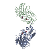
| ||||||||
|---|---|---|---|---|---|---|---|---|---|
| 1 |
| ||||||||
| Unit cell |
| ||||||||
| Noncrystallographic symmetry (NCS) | NCS oper: (Code: given Matrix: (-0.99956, -0.01683, 0.02444), Vector : |
-Components
-Protein , 1 types, 2 molecules AB
| #1: Protein | Mass: 28904.062 Da / Num. of mol.: 2 Source method: isolated from a genetically manipulated source Source: (gene. exp.) Homo sapiens (human) / Tissue: LIVER / Description: HETEROLOGOUSLY EXPRESSED / Plasmid: PKK 223-3,ACCI-DELETED / Production host:  Escherichia coli (E. coli) / Strain (production host): JM 109 Escherichia coli (E. coli) / Strain (production host): JM 109References: UniProt: Q16775, hydroxyacylglutathione hydrolase |
|---|
-Non-polymers , 6 types, 382 molecules 


| #2: Chemical | ChemComp-ZN /  Mass: 65.409 Da / Num. of mol.: 4 / Source method: obtained synthetically / Formula: Zn Mass: 65.409 Da / Num. of mol.: 4 / Source method: obtained synthetically / Formula: Zn#3: Chemical | ChemComp-CAC / Cacodylic acid Mass: 136.989 Da / Num. of mol.: 4 / Source method: obtained synthetically / Formula: C2H6AsO2 Mass: 136.989 Da / Num. of mol.: 4 / Source method: obtained synthetically / Formula: C2H6AsO2#4: Chemical | Acetate Mass: 59.044 Da / Num. of mol.: 2 / Source method: obtained synthetically / Formula: C2H3O2 Mass: 59.044 Da / Num. of mol.: 2 / Source method: obtained synthetically / Formula: C2H3O2#5: Chemical |  Mass: 54.938 Da / Num. of mol.: 2 / Source method: obtained synthetically / Formula: Mn Mass: 54.938 Da / Num. of mol.: 2 / Source method: obtained synthetically / Formula: Mn#6: Chemical | ChemComp-CL / | Chloride Mass: 35.453 Da / Num. of mol.: 1 / Source method: obtained synthetically / Formula: Cl Mass: 35.453 Da / Num. of mol.: 1 / Source method: obtained synthetically / Formula: Cl#7: Water | ChemComp-HOH / | WaterMass: 18.015 Da / Num. of mol.: 369 / Source method: isolated from a natural source / Formula: H2O |
|---|
-Details
| Nonpolymer details | ONLY THE ARSENIC ATOMS OF CAC A265 AND CAC B265 HAVE BEEN MODELLED. CL 467 TENTATIVELY MODELLED AS ...ONLY THE ARSENIC ATOMS OF CAC A265 AND CAC B265 HAVE BEEN MODELLED. CL 467 TENTATIVEL |
|---|
-Experimental details
-Experiment
| Experiment | Method: X-RAY DIFFRACTION / Number of used crystals: 1 |
|---|
- Sample preparation
| Crystal | Density Matthews: 2 Å3/Da / Density % sol: 29 % | ||||||||||||||||||||||||||||||||||||||||||||||||||||||
|---|---|---|---|---|---|---|---|---|---|---|---|---|---|---|---|---|---|---|---|---|---|---|---|---|---|---|---|---|---|---|---|---|---|---|---|---|---|---|---|---|---|---|---|---|---|---|---|---|---|---|---|---|---|---|---|
| Crystal grow | pH: 6.5 Details: EQUILABRATION AGAINST 15-30% W/V PEG 2000 MONOMETHYL ETHER 0.2M MG-ACETATE, 0.1M NA-CACODYLATE PH 6.5 AND 2MM DTT | ||||||||||||||||||||||||||||||||||||||||||||||||||||||
| Crystal | *PLUS | ||||||||||||||||||||||||||||||||||||||||||||||||||||||
| Crystal grow | *PLUS pH: 7.1 / Method: vapor diffusion, hanging drop | ||||||||||||||||||||||||||||||||||||||||||||||||||||||
| Components of the solutions | *PLUS
|
-Data collection
| Diffraction | Mean temperature: 100 K |
|---|---|
| Diffraction source | Source: SYNCHROTRON / Site: EMBL/DESY, HAMBURG  / Beamline: X11 / Wavelength: 0.911 / Beamline: X11 / Wavelength: 0.911 |
| Detector | Type: MARRESEARCH / Detector: IMAGE PLATE / Date: Nov 1, 1997 / Details: BENT MIRROR |
| Radiation | Monochromator: TRIANGULAR / Protocol: SINGLE WAVELENGTH / Monochromatic (M) / Laue (L): M / Scattering type: x-ray |
| Radiation wavelength | Wavelength: 0.911 Å / Relative weight: 1 |
| Reflection | Resolution: 1.9→15 Å / Num. obs: 126060 / % possible obs: 99 % / Redundancy: 3.4 % / Biso Wilson estimate: 18.3 Å2 / Rmerge(I) obs: 0.063 / Net I/σ(I): 14 |
| Reflection shell | Resolution: 1.9→1.93 Å / Redundancy: 2.7 % / Rmerge(I) obs: 0.31 / Mean I/σ(I) obs: 4 / % possible all: 99 |
| Reflection | *PLUS Num. obs: 36838 / Num. measured all: 126060 |
| Reflection shell | *PLUS % possible obs: 99 % / Rmerge(I) obs: 0.312 |
- Processing
| Software |
| ||||||||||||||||||||||||||||||||||||||||||||||||||||||||||||||||||||||||||||||||||||
|---|---|---|---|---|---|---|---|---|---|---|---|---|---|---|---|---|---|---|---|---|---|---|---|---|---|---|---|---|---|---|---|---|---|---|---|---|---|---|---|---|---|---|---|---|---|---|---|---|---|---|---|---|---|---|---|---|---|---|---|---|---|---|---|---|---|---|---|---|---|---|---|---|---|---|---|---|---|---|---|---|---|---|---|---|---|
| Refinement | Method to determine structure: SIRAS / Resolution: 1.9→15 Å / SU B: 3.6 / SU ML: 0.11 / Cross valid method: THROUGHOUT / σ(F): 0 / ESU R: 0.18 / ESU R Free: 0.17 / Details: VAN DER WAALS RADII ON ZINC IONS SET TO 0.1A
| ||||||||||||||||||||||||||||||||||||||||||||||||||||||||||||||||||||||||||||||||||||
| Displacement parameters | Biso mean: 20.8 Å2 | ||||||||||||||||||||||||||||||||||||||||||||||||||||||||||||||||||||||||||||||||||||
| Refinement step | Cycle: LAST / Resolution: 1.9→15 Å
| ||||||||||||||||||||||||||||||||||||||||||||||||||||||||||||||||||||||||||||||||||||
| Refine LS restraints |
| ||||||||||||||||||||||||||||||||||||||||||||||||||||||||||||||||||||||||||||||||||||
| Software | *PLUS Name: REFMAC / Classification: refinement | ||||||||||||||||||||||||||||||||||||||||||||||||||||||||||||||||||||||||||||||||||||
| Refinement | *PLUS Highest resolution: 1.9 Å / σ(F): 0 / % reflection Rfree: 5 % | ||||||||||||||||||||||||||||||||||||||||||||||||||||||||||||||||||||||||||||||||||||
| Solvent computation | *PLUS | ||||||||||||||||||||||||||||||||||||||||||||||||||||||||||||||||||||||||||||||||||||
| Displacement parameters | *PLUS Biso mean: 20.8 Å2 | ||||||||||||||||||||||||||||||||||||||||||||||||||||||||||||||||||||||||||||||||||||
| Refine LS restraints | *PLUS
|
