 Movie
Movie Controller

[English] 日本語
 Yorodumi
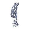
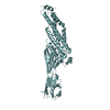
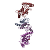
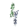
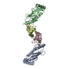
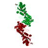
- PDB-1qfh: DIMERIZATION OF GELATION FACTOR FROM DICTYOSTELIUM DISCOIDEUM: CR... -
+ Open data

- Basic information
| Entry | Database: PDB / ID: 1qfh | ||||||
|---|---|---|---|---|---|---|---|
| Title | DIMERIZATION OF GELATION FACTOR FROM DICTYOSTELIUM DISCOIDEUM: CRYSTAL STRUCTURE OF ROD DOMAINS 5 AND 6 | ||||||
 Components Components | PROTEIN (GELATION FACTOR) | ||||||
 Keywords Keywords | ACTIN BINDING PROTEIN /  IMMUNOGLOBULIN / GELATION FACTOR / ABP-120 IMMUNOGLOBULIN / GELATION FACTOR / ABP-120 | ||||||
| Function / homology |  Function and homology informationregulation of pseudopodium assembly / anterior cell cortex / pseudopodium assembly / sorocarp development / posterior cell cortex / chemotaxis to cAMP / lateral cell cortex / phototaxis / macropinocytic cup / protein kinase B binding ...regulation of pseudopodium assembly / anterior cell cortex / pseudopodium assembly / sorocarp development / posterior cell cortex / chemotaxis to cAMP / lateral cell cortex / phototaxis / macropinocytic cup / protein kinase B binding / actin crosslink formation / thermotaxis / hyperosmotic response / mitogen-activated protein kinase binding / lamellipodium assembly / phagocytic cup / cortical actin cytoskeleton / cell leading edge / pseudopodium / phagocytosis / response to cAMP / extracellular matrix / cell motility / small GTPase binding / actin filament binding / cell migration / cell cortex / actin cytoskeleton organization / plasma membrane / cytoplasm Function and homology informationregulation of pseudopodium assembly / anterior cell cortex / pseudopodium assembly / sorocarp development / posterior cell cortex / chemotaxis to cAMP / lateral cell cortex / phototaxis / macropinocytic cup / protein kinase B binding ...regulation of pseudopodium assembly / anterior cell cortex / pseudopodium assembly / sorocarp development / posterior cell cortex / chemotaxis to cAMP / lateral cell cortex / phototaxis / macropinocytic cup / protein kinase B binding / actin crosslink formation / thermotaxis / hyperosmotic response / mitogen-activated protein kinase binding / lamellipodium assembly / phagocytic cup / cortical actin cytoskeleton / cell leading edge / pseudopodium / phagocytosis / response to cAMP / extracellular matrix / cell motility / small GTPase binding / actin filament binding / cell migration / cell cortex / actin cytoskeleton organization / plasma membrane / cytoplasmSimilarity search - Function | ||||||
| Biological species |  Dictyostelium discoideum (eukaryote) Dictyostelium discoideum (eukaryote) | ||||||
| Method | X-RAY DIFFRACTION / SYNCHROTRON / MAD / Resolution: 2.2 Å | ||||||
 Authors Authors | Mccoy, A.J. / Fucini, P. / Noegel, A.A. / Stewart, M. | ||||||
 Citation Citation | Journal: Nat.Struct.Biol. / Year: 1999 Title: Structural basis for dimerization of the Dictyostelium gelation factor (ABP120) rod. Authors: McCoy, A.J. / Fucini, P. / Noegel, A.A. / Stewart, M. #1: Journal: J.Struct.Biol. / Year: 1997 Title: Crystallization and preliminary X-Ray diffraction characterization of a dimerizing fragment of the rod domain of the Dictyostelium gelation factor (ABP-120). Authors: Fucini, P. / McCoy, A.J. / Gomez-Ortiz, M. / Schleicher, M. / Noegel, A.A. / Stewart, M. | ||||||
| History |
|
- Structure visualization
| Structure viewer | Molecule: MolmilJmol/JSmol |
|---|
- Downloads & links
-Download
| PDBx/mmCIF format | 1qfh.cif.gz | 93 KB | Display | PDBx/mmCIF format |
|---|---|---|---|---|
| PDB format | pdb1qfh.ent.gz | 71.1 KB | Display | PDB format |
| PDBx/mmJSON format | 1qfh.json.gz | Tree view | PDBx/mmJSON format | |
| Others |  Other downloads Other downloads |
-Validation report
| Arichive directory | https://data.pdbj.org/pub/pdb/validation_reports/qf/1qfhftp://data.pdbj.org/pub/pdb/validation_reports/qf/1qfh | HTTPS FTP |
|---|
-Related structure data
| Similar structure data |
|---|


-Links
 PDBj
PDBj- Assembly
| Deposited unit | 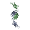
| ||||||||||||
|---|---|---|---|---|---|---|---|---|---|---|---|---|---|
| 1 |
| ||||||||||||
| Unit cell |
| ||||||||||||
| Noncrystallographic symmetry (NCS) | NCS oper:
|
-Components
| #1: Protein | Mass: 22355.594 Da / Num. of mol.: 2 / Fragment: ROD DOMAINS 5 AMD 6 Source method: isolated from a genetically manipulated source Source: (gene. exp.) Dictyostelium discoideum (eukaryote) / Production host:  Escherichia coli (E. coli) / References: UniProt: P13466 Escherichia coli (E. coli) / References: UniProt: P13466#2: Water | ChemComp-HOH / | Water Mass: 18.015 Da / Num. of mol.: 237 / Source method: isolated from a natural source / Formula: H2O Mass: 18.015 Da / Num. of mol.: 237 / Source method: isolated from a natural source / Formula: H2O |
|---|
-Experimental details
-Experiment
| Experiment | Method: X-RAY DIFFRACTION / Number of used crystals: 1 |
|---|
- Sample preparation
| Crystal | Density Matthews: 3.1 Å3/Da / Density % sol: 40 % | ||||||||||||||||||||||||
|---|---|---|---|---|---|---|---|---|---|---|---|---|---|---|---|---|---|---|---|---|---|---|---|---|---|
| Crystal grow | pH: 7.2 / Details: 30% PEG 1000, 0.1M TRIS_HCL PH 7.2, 25% GLYCEROL | ||||||||||||||||||||||||
| Crystal | *PLUS | ||||||||||||||||||||||||
| Crystal grow | *PLUS Temperature: 20 ℃ / pH: 7.4 / Method: unknown | ||||||||||||||||||||||||
| Components of the solutions | *PLUS
|
-Data collection
| Diffraction | Mean temperature: 100 K |
|---|---|
| Diffraction source | Source: SYNCHROTRON / Site: SRS  / Beamline: PX9.6 / Wavelength: 0.88 / Beamline: PX9.6 / Wavelength: 0.88 |
| Detector | Type: MARRESEARCH / Detector: IMAGE PLATE / Date: Jun 1, 1997 |
| Radiation | Protocol: SINGLE WAVELENGTH / Monochromatic (M) / Laue (L): M / Scattering type: x-ray |
| Radiation wavelength | Wavelength: 0.88 Å / Relative weight: 1 |
| Reflection | Resolution: 2.2→26.23 Å / Num. obs: 82513 / % possible obs: 98 % / Redundancy: 2.9 % / Biso Wilson estimate: 29.87 Å2 / Rmerge(I) obs: 0.048 / Net I/σ(I): 8.8 |
| Reflection shell | Resolution: 2.2→2.32 Å / Redundancy: 2.9 % / Rmerge(I) obs: 0.091 / Mean I/σ(I) obs: 6.9 / % possible all: 97.6 |
| Reflection | *PLUS Num. obs: 28551 / % possible obs: 98 % / Num. measured all: 82513 |
| Reflection shell | *PLUS % possible obs: 97.6 % / Num. unique obs: 4076 / Num. measured obs: 11993 |
- Processing
| Software |
| ||||||||||||||||||||||||||||||||||||||||||||||||||||||||||||||||||||||||||||||||||||
|---|---|---|---|---|---|---|---|---|---|---|---|---|---|---|---|---|---|---|---|---|---|---|---|---|---|---|---|---|---|---|---|---|---|---|---|---|---|---|---|---|---|---|---|---|---|---|---|---|---|---|---|---|---|---|---|---|---|---|---|---|---|---|---|---|---|---|---|---|---|---|---|---|---|---|---|---|---|---|---|---|---|---|---|---|---|
| Refinement | Method to determine structure: MAD / Resolution: 2.2→26.63 Å / SU B: 5.42051 / SU ML: 0.14239 / Cross valid method: THROUGHOUT / σ(F): 0 / ESU R: 0.24148 / ESU R Free: 0.20983
| ||||||||||||||||||||||||||||||||||||||||||||||||||||||||||||||||||||||||||||||||||||
| Displacement parameters | Biso mean: 42.59 Å2 | ||||||||||||||||||||||||||||||||||||||||||||||||||||||||||||||||||||||||||||||||||||
| Refinement step | Cycle: LAST / Resolution: 2.2→26.63 Å
| ||||||||||||||||||||||||||||||||||||||||||||||||||||||||||||||||||||||||||||||||||||
| Refine LS restraints |
| ||||||||||||||||||||||||||||||||||||||||||||||||||||||||||||||||||||||||||||||||||||
| Software | *PLUS Name: REFMAC / Classification: refinement | ||||||||||||||||||||||||||||||||||||||||||||||||||||||||||||||||||||||||||||||||||||
| Refinement | *PLUS Highest resolution: 2.2 Å / σ(F): 0 / % reflection Rfree: 5 % | ||||||||||||||||||||||||||||||||||||||||||||||||||||||||||||||||||||||||||||||||||||
| Solvent computation | *PLUS | ||||||||||||||||||||||||||||||||||||||||||||||||||||||||||||||||||||||||||||||||||||
| Displacement parameters | *PLUS |
