 Movie
Movie Controller

[English] 日本語
 Yorodumi
- PDB-1qcz: CRYSTAL STRUCTURE OF E. COLI PURE, AN UNUSUAL MUTASE THAT CATALYZ... -
+ Open data

- Basic information
| Entry | Database: PDB / ID: 1qcz | ||||||
|---|---|---|---|---|---|---|---|
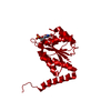
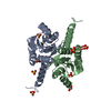
| Title | CRYSTAL STRUCTURE OF E. COLI PURE, AN UNUSUAL MUTASE THAT CATALYZES THE CONVERSION OF N5-CARBOXYAMINOIMIDAZOLE RIBONUCLEOTIDE (N5-CAIR) TO 4-CARBOXYAMINOIMIDAZOLE RIBONUCLEOTIDE (CAIR) IN THE PURINE BIOSYNTHETIC PATHWAY | ||||||
 Components Components | N5-CARBOXYAMINOIMIDAZOLE RIBONUCLEOTIDE MUTASE | ||||||
 Keywords Keywords |  LYASE / THREE-LAYER (ALPHA-BETA-ALPHA) SANDWICH LYASE / THREE-LAYER (ALPHA-BETA-ALPHA) SANDWICH | ||||||
| Function / homology |  Function and homology information5-(carboxyamino)imidazole ribonucleotide mutase / 5-(carboxyamino)imidazole ribonucleotide mutase activity / 'de novo' IMP biosynthetic process / identical protein binding / cytosol Function and homology information5-(carboxyamino)imidazole ribonucleotide mutase / 5-(carboxyamino)imidazole ribonucleotide mutase activity / 'de novo' IMP biosynthetic process / identical protein binding / cytosolSimilarity search - Function | ||||||
| Biological species |  Escherichia coli (E. coli) Escherichia coli (E. coli) | ||||||
| Method | X-RAY DIFFRACTION / SYNCHROTRON / MAD / Resolution: 1.5 Å | ||||||
 Authors Authors | Ealick, S.E. / Mathews, I.I. | ||||||
 Citation Citation | Journal: Structure Fold.Des. / Year: 1999 Title: Crystal structure of Escherichia coli PurE, an unusual mutase in the purine biosynthetic pathway. Authors: Mathews, I.I. / Kappock, T.J. / Stubbe, J. / Ealick, S.E. #1: Journal: Biochemistry / Year: 1999Title: Evidence for the Direct Transfer of the Carboxylate of N5-Carboxyaminoimidazole Ribonucleotide (N5-CAIR) to Generate 4-Carboxy-5-Aminoimidazole Ribonucleotide Catalyzed by Escherichia coli purE, an N5-CAIR Mutase Authors: Meyer, E. / Kappock, T.J. / Osuji, C. / Stubbe, J. #2: Journal: Biochemistry / Year: 1994Title: Reactions Catalyzed by 5-Aminoimidazole Ribonucleotide Carboxylases from Escherichia coli Carboxylases from Escherichia coli and Gallus gallus: a Case for Divergent Catalytic Mechanisms Authors: Firestine, S.M. / Poon, S.W. / Mueller, E.J. / Stubbe, J. / Davisson, V.J. #3: Journal: Biochemistry / Year: 1994Title: N5-Carboxyaminoimidazole Ribonucleotide: Evidence for a New Intermediate and Two New Enzymatic Activities in the de novo Purine Biosynthetic Pathway of Escherichia coli Authors: Mueller, E.J. / Meyer, E. / Rudolph, J. / Davisson, V.J. / Stubbe, J. #4: Journal: J.Bacteriol. / Year: 1989Title: Nucleotide Sequence Analysis of the purEK Operon Encoding 5'-Phosphoribosyl-5-Aminoimidazole Carboxylase of Escherichia coli K-12 Authors: Tiedeman, A.A. / Keyhani, J. / Kamholz, J. / 3D Daum, H.A. / Gots, J.S. / Smith, J.M. #5: Journal: J.Bacteriol. / Year: 1989Title: Identification and Sequence Analysis of Escherichia coli purE and purK Genes Encoding 5'-Phosphoribosyl-5-Amino-4-Imidazole Carboxylase for de novo Purine Biosynthesis Authors: Watanabe, W. / Sampei, G. / Aiba, A. / Mizobuchi, K. | ||||||
| History |
|
- Structure visualization
| Structure viewer | Molecule: MolmilJmol/JSmol |
|---|
- Downloads & links
-Download
| PDBx/mmCIF format | 1qcz.cif.gz | 44.3 KB | Display | PDBx/mmCIF format |
|---|---|---|---|---|
| PDB format | pdb1qcz.ent.gz | 33.6 KB | Display | PDB format |
| PDBx/mmJSON format | 1qcz.json.gz | Tree view | PDBx/mmJSON format | |
| Others |  Other downloads Other downloads |
-Validation report
| Arichive directory | https://data.pdbj.org/pub/pdb/validation_reports/qc/1qczftp://data.pdbj.org/pub/pdb/validation_reports/qc/1qcz | HTTPS FTP |
|---|
-Related structure data


-Links
 PDBj
PDBj- Assembly
| Deposited unit | 
| |||||||||||||||||||||
|---|---|---|---|---|---|---|---|---|---|---|---|---|---|---|---|---|---|---|---|---|---|---|
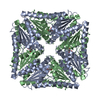
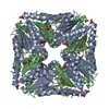
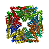
| 1 | x 8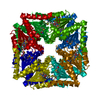
| |||||||||||||||||||||
| Unit cell |
| |||||||||||||||||||||
| Components on special symmetry positions |
| |||||||||||||||||||||
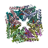
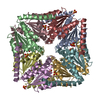
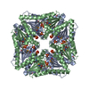
| Details | The biological assembly is a octamer which is generated from chain A by the 4-fold and 2-fold symmetry |
-Components
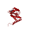
| #1: Protein | Mass: 17986.852 Da / Num. of mol.: 1 Source method: isolated from a genetically manipulated source Source: (gene. exp.) Escherichia coli (E. coli) / Plasmid: PNC2 / Gene (production host): PURE / Production host: Escherichia coli (E. coli) / Strain (production host): B834(DE3)References: UniProt: P09028, UniProt: P0AG18*PLUS, phosphoribosylaminoimidazole carboxylase |
|---|---|
| #2: Water | ChemComp-HOH / Water Mass: 18.015 Da / Num. of mol.: 171 / Source method: isolated from a natural source / Formula: H2O Mass: 18.015 Da / Num. of mol.: 171 / Source method: isolated from a natural source / Formula: H2O |
| Compound details | THE SUBSTRATE SPECIFICITY OF E. COLI PURE FOR N5-CAIR DIFFERS FROM VERTEBRATE PURE (AIR ...THE SUBSTRATE SPECIFICIT |
-Experimental details
-Experiment
| Experiment | Method: X-RAY DIFFRACTION / Number of used crystals: 1 |
|---|
- Sample preparation
| Crystal | Density Matthews: 2.22 Å3/Da / Density % sol: 55 % | ||||||||||||||||||||||||
|---|---|---|---|---|---|---|---|---|---|---|---|---|---|---|---|---|---|---|---|---|---|---|---|---|---|
| Crystal grow | Temperature: 291 K / Method: vapor diffusion, hanging drop / pH: 8 Details: PEG400, TRIS.HCL, MAGNESIUM CHLORIDE, pH 8.00, VAPOR DIFFUSION, HANGING DROP, temperature 18K | ||||||||||||||||||||||||
| Crystal grow | *PLUS Details: drop consists of 1:1 mixture of well and protein solutions | ||||||||||||||||||||||||
| Components of the solutions | *PLUS
|
-Data collection
| Diffraction | Mean temperature: 180 K |
|---|---|
| Diffraction source | Source: SYNCHROTRON / Site: CHESS  / Beamline: F1 / Wavelength: 0.918 / Beamline: F1 / Wavelength: 0.918 |
| Detector | Type: ADSC QUANTUM 4 / Detector: CCD / Date: Jan 1, 1998 |
| Radiation | Protocol: SINGLE WAVELENGTH / Monochromatic (M) / Laue (L): M / Scattering type: x-ray |
| Radiation wavelength | Wavelength: 0.918 Å / Relative weight: 1 |
| Reflection | Resolution: 1.5→20 Å / Num. all: 193682 / Num. obs: 25072 / % possible obs: 97.2 % / Observed criterion σ(I): 1 / Redundancy: 7.7 % / Biso Wilson estimate: 14.6 Å2 / Rmerge(I) obs: 0.077 / Net I/σ(I): 6.8 |
| Reflection shell | Resolution: 1.5→1.58 Å / Redundancy: 3.9 % / Rmerge(I) obs: 0.172 / % possible all: 86.7 |
| Reflection | *PLUS Num. measured all: 193682 |
| Reflection shell | *PLUS % possible obs: 86.7 % |
- Processing
| Software |
| ||||||||||||||||||||||||||||||||||||||||||||||||||||||||||||||||||||||||||||||||
|---|---|---|---|---|---|---|---|---|---|---|---|---|---|---|---|---|---|---|---|---|---|---|---|---|---|---|---|---|---|---|---|---|---|---|---|---|---|---|---|---|---|---|---|---|---|---|---|---|---|---|---|---|---|---|---|---|---|---|---|---|---|---|---|---|---|---|---|---|---|---|---|---|---|---|---|---|---|---|---|---|---|
| Refinement | Method to determine structure: MAD / Resolution: 1.5→20 Å / σ(F): 2 / σ(I): 1 / Stereochemistry target values: ENGH & HUBER
| ||||||||||||||||||||||||||||||||||||||||||||||||||||||||||||||||||||||||||||||||
| Displacement parameters | Biso mean: 14.2 Å2 | ||||||||||||||||||||||||||||||||||||||||||||||||||||||||||||||||||||||||||||||||
| Refine analyze |
| ||||||||||||||||||||||||||||||||||||||||||||||||||||||||||||||||||||||||||||||||
| Refinement step | Cycle: LAST / Resolution: 1.5→20 Å
| ||||||||||||||||||||||||||||||||||||||||||||||||||||||||||||||||||||||||||||||||
| Refine LS restraints |
| ||||||||||||||||||||||||||||||||||||||||||||||||||||||||||||||||||||||||||||||||
| LS refinement shell | Resolution: 1.5→1.57 Å / Total num. of bins used: 8
| ||||||||||||||||||||||||||||||||||||||||||||||||||||||||||||||||||||||||||||||||
| Software | *PLUS Name: X-PLOR / Version: 3.843 / Classification: refinement | ||||||||||||||||||||||||||||||||||||||||||||||||||||||||||||||||||||||||||||||||
| Refine LS restraints | *PLUS
|
