 Movie
Movie Controller


+ Open data

- Basic information
| Entry | Database: PDB / ID: 1q6x | ||||||
|---|---|---|---|---|---|---|---|
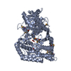
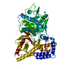
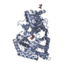
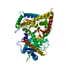
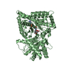
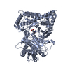
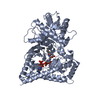
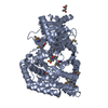
| Title | Crystal structure of rat choline acetyltransferase | ||||||
 Components Components | choline O-acetyltransferase Choline acetyltransferase Choline acetyltransferase | ||||||
 Keywords Keywords | TRANSFERASE / alpha beta sandwich / extended loop / two domains | ||||||
| Function / homology |  Function and homology informationcholine O-acetyltransferase / choline O-acetyltransferase activity / Synthesis of PC / acetylcholine biosynthetic process / rhythmic excitation / establishment of synaptic specificity at neuromuscular junction / rhythmic behavior / Acetylcholine Neurotransmitter Release Cycle / neuromuscular synaptic transmission / choline binding ...choline O-acetyltransferase / choline O-acetyltransferase activity / Synthesis of PC / acetylcholine biosynthetic process / rhythmic excitation / establishment of synaptic specificity at neuromuscular junction / rhythmic behavior / Acetylcholine Neurotransmitter Release Cycle / neuromuscular synaptic transmission / choline binding / adult walking behavior / muscle organ development / antral ovarian follicle growth / dendrite development / response to nutrient / memory / neuron differentiation / chemical synaptic transmission / response to ethanol / response to hypoxia / neuron projection / response to xenobiotic stimulus / axon / neuronal cell body / synapse / cytoplasm Function and homology informationcholine O-acetyltransferase / choline O-acetyltransferase activity / Synthesis of PC / acetylcholine biosynthetic process / rhythmic excitation / establishment of synaptic specificity at neuromuscular junction / rhythmic behavior / Acetylcholine Neurotransmitter Release Cycle / neuromuscular synaptic transmission / choline binding ...choline O-acetyltransferase / choline O-acetyltransferase activity / Synthesis of PC / acetylcholine biosynthetic process / rhythmic excitation / establishment of synaptic specificity at neuromuscular junction / rhythmic behavior / Acetylcholine Neurotransmitter Release Cycle / neuromuscular synaptic transmission / choline binding / adult walking behavior / muscle organ development / antral ovarian follicle growth / dendrite development / response to nutrient / memory / neuron differentiation / chemical synaptic transmission / response to ethanol / response to hypoxia / neuron projection / response to xenobiotic stimulus / axon / neuronal cell body / synapse / cytoplasmSimilarity search - Function | ||||||
| Biological species |  Rattus norvegicus (Norway rat) Rattus norvegicus (Norway rat) | ||||||
| Method | X-RAY DIFFRACTION / SYNCHROTRON / MOLECULAR REPLACEMENT / Resolution: 2.5 Å | ||||||
 Authors Authors | Cai, Y. / Rodgers, D.W. | ||||||
 Citation Citation | Journal: Embo J. / Year: 2004 Title: Choline acetyltransferase structure reveals distribution of mutations that cause motor disorders. Authors: Cai, Y. / Cronin, C.N. / Engel, A.G. / Ohno, K. / Hersh, L.B. / Rodgers, D.W. | ||||||
| History |
| ||||||
| Remark 999 | SEQUENCE THE AUTHOR MAINTAINS THAT THE SEQUENCE IN THE SEQUENCE DATABASE IS INCORRECT |
- Structure visualization
| Structure viewer | Molecule: MolmilJmol/JSmol |
|---|
- Downloads & links
-Download
| PDBx/mmCIF format | 1q6x.cif.gz | 248.8 KB | Display | PDBx/mmCIF format |
|---|---|---|---|---|
| PDB format | pdb1q6x.ent.gz | 198.2 KB | Display | PDB format |
| PDBx/mmJSON format | 1q6x.json.gz | Tree view | PDBx/mmJSON format | |
| Others |  Other downloads Other downloads |
-Validation report
| Arichive directory | https://data.pdbj.org/pub/pdb/validation_reports/q6/1q6xftp://data.pdbj.org/pub/pdb/validation_reports/q6/1q6x | HTTPS FTP |
|---|
-Related structure data
| Related structure data |  1ndbS S: Starting model for refinement |
|---|---|
| Similar structure data |


-Links
 PDBj
PDBj


- Assembly
| Deposited unit | 
| ||||||||
|---|---|---|---|---|---|---|---|---|---|
| 1 | 
| ||||||||
| 2 | 
| ||||||||
| Unit cell |
| ||||||||
| Details | The biological assembly is a single polypeptide chain, and there are two biological assemblies in the asymmetric unit. |
-Components
| #1: Protein | Choline acetyltransferase Mass: 72475.164 Da / Num. of mol.: 2 Source method: isolated from a genetically manipulated source Source: (gene. exp.) Rattus norvegicus (Norway rat) / Gene: cholinergic gene locus / Plasmid: pLENTY / Production host:  Escherichia coli (E. coli) / Strain (production host): DH5alphaF'IQ / References: UniProt: P32738, choline O-acetyltransferase Escherichia coli (E. coli) / Strain (production host): DH5alphaF'IQ / References: UniProt: P32738, choline O-acetyltransferase#2: Chemical |   Mass: 22.990 Da / Num. of mol.: 2 / Source method: obtained synthetically / Formula: Na Mass: 22.990 Da / Num. of mol.: 2 / Source method: obtained synthetically / Formula: Na#3: Water | ChemComp-HOH / | Water Mass: 18.015 Da / Num. of mol.: 345 / Source method: isolated from a natural source / Formula: H2O Mass: 18.015 Da / Num. of mol.: 345 / Source method: isolated from a natural source / Formula: H2O |
|---|
-Experimental details
-Experiment
| Experiment | Method: X-RAY DIFFRACTION / Number of used crystals: 1 |
|---|
- Sample preparation
| Crystal | Density Matthews: 3.18 Å3/Da / Density % sol: 61.27 % |
|---|---|
| Crystal grow | Temperature: 277 K / Method: vapor diffusion, sitting drop / pH: 6.5 Details: PEG 20000, (2-(N-Morpholino)ethanesulfonic acid, sodium chloride, 2-mercaptoethanol, pH 6.5, VAPOR DIFFUSION, SITTING DROP, temperature 277K |
-Data collection
| Diffraction | Mean temperature: 110 K |
|---|---|
| Diffraction source | Source: SYNCHROTRON / Site: APS  / Beamline: 22-ID / Wavelength: 0.97934 Å / Beamline: 22-ID / Wavelength: 0.97934 Å |
| Detector | Type: MARRESEARCH / Detector: CCD / Date: Nov 23, 2002 / Details: double crystal focusing mirror |
| Radiation | Monochromator: double crystal / Protocol: SINGLE WAVELENGTH / Monochromatic (M) / Laue (L): M / Scattering type: x-ray |
| Radiation wavelength | Wavelength: 0.97934 Å / Relative weight: 1 |
| Reflection | Resolution: 2.5→30 Å / Num. all: 62300 / Num. obs: 62219 / Observed criterion σ(F): -4 / Observed criterion σ(I): -4 / Redundancy: 3.8 % / Biso Wilson estimate: 38.3 Å2 / Rmerge(I) obs: 0.075 / Net I/σ(I): 8.1 |
| Reflection shell | Resolution: 2.5→2.59 Å / Redundancy: 3.76 % / Rmerge(I) obs: 0.337 / Mean I/σ(I) obs: 3.9 / Num. unique all: 6012 / % possible all: 95.2 |
- Processing
| Software |
| ||||||||||||||||||||||||||||||||||||
|---|---|---|---|---|---|---|---|---|---|---|---|---|---|---|---|---|---|---|---|---|---|---|---|---|---|---|---|---|---|---|---|---|---|---|---|---|---|
| Refinement | Method to determine structure: MOLECULAR REPLACEMENT Starting model: PDB entry 1NDB Resolution: 2.5→28.94 Å / Rfactor Rfree error: 0.003 / Isotropic thermal model: RESTRAINED / Cross valid method: THROUGHOUT / σ(F): 0 / Stereochemistry target values: Engh & Huber Details: Only C-alpha positions could be assigned for residues 356-367 in both chains A and B
| ||||||||||||||||||||||||||||||||||||
| Solvent computation | Solvent model: FLAT MODEL / Bsol: 16.3927 Å2 / ksol: 0.312752 e/Å3 | ||||||||||||||||||||||||||||||||||||
| Displacement parameters | Biso mean: 30.7 Å2
| ||||||||||||||||||||||||||||||||||||
| Refine analyze |
| ||||||||||||||||||||||||||||||||||||
| Refinement step | Cycle: LAST / Resolution: 2.5→28.94 Å
| ||||||||||||||||||||||||||||||||||||
| Refine LS restraints |
| ||||||||||||||||||||||||||||||||||||
| LS refinement shell | Resolution: 2.5→2.66 Å / Rfactor Rfree error: 0.01 / Total num. of bins used: 6
| ||||||||||||||||||||||||||||||||||||
| Xplor file |
|
