 Movie
Movie Controller

[English] 日本語
 Yorodumi
- PDB-1pzu: An asymmetric NFAT1-RHR homodimer on a pseudo-palindromic, Kappa-... -
+ Open data

- Basic information
| Entry | Database: PDB / ID: 1pzu | ||||||
|---|---|---|---|---|---|---|---|
| Title | An asymmetric NFAT1-RHR homodimer on a pseudo-palindromic, Kappa-B site | ||||||
 Components Components |
| ||||||
 Keywords Keywords | TRANSCRIPTION/DNA /  TRANSCRIPTION FACTOR / NFAT1-RHR homodimer / PROTEIN-DNA COMPLEX / human IL8 promoter / TRANSCRIPTION-DNA COMPLEX TRANSCRIPTION FACTOR / NFAT1-RHR homodimer / PROTEIN-DNA COMPLEX / human IL8 promoter / TRANSCRIPTION-DNA COMPLEX | ||||||
| Function / homology |  Function and homology information Function and homology information: / transcription factor AP-1 complex / negative regulation of vascular associated smooth muscle cell differentiation / myotube cell development / RUNX1 and FOXP3 control the development of regulatory T lymphocytes (Tregs) / calcineurin-NFAT signaling cascade / cartilage development / CLEC7A (Dectin-1) induces NFAT activation / positive regulation of myoblast fusion / Calcineurin activates NFAT ...: / transcription factor AP-1 complex / negative regulation of vascular associated smooth muscle cell differentiation / myotube cell development / RUNX1 and FOXP3 control the development of regulatory T lymphocytes (Tregs) / calcineurin-NFAT signaling cascade / cartilage development / CLEC7A (Dectin-1) induces NFAT activation / positive regulation of myoblast fusion / Calcineurin activates NFAT / phosphatase binding / positive regulation of B cell proliferation / cellular response to calcium ion / FCERI mediated Ca+2 mobilization / 14-3-3 protein binding / B cell receptor signaling pathway / DNA-binding transcription repressor activity, RNA polymerase II-specific / cell migration / sequence-specific double-stranded DNA binding / DNA-binding transcription activator activity, RNA polymerase II-specific / transcription regulator complex / transcription by RNA polymerase II / molecular adaptor activity / DNA-binding transcription factor activity, RNA polymerase II-specific / response to xenobiotic stimulus / ribonucleoprotein complex / RNA polymerase II cis-regulatory region sequence-specific DNA binding / DNA-binding transcription factor activity / DNA damage response / chromatin binding / chromatin / positive regulation of gene expression / regulation of DNA-templated transcription / regulation of transcription by RNA polymerase II / positive regulation of DNA-templated transcription / DNA binding / nucleoplasm / nucleus / cytosol / cytoplasmSimilarity search - Function | ||||||
| Biological species |  Homo sapiens (human) Homo sapiens (human) | ||||||
| Method | X-RAY DIFFRACTION / SYNCHROTRON / MOLECULAR REPLACEMENT / Resolution: 3.1 Å | ||||||
 Authors Authors | Jin, L. / Sliz, P. / Chen, L. / Macian, F. / Rao, A. / Hogan, P.G. / Harrison, S.C. | ||||||
 Citation Citation | Journal: Nat.Struct.Biol. / Year: 2003 Title: An asymmetric NFAT1 dimer on a pseudo-palindromic KB-like DNA site Authors: Jin, L. / Sliz, P. / Chen, L. / Macian, F. / Rao, A. / Hogan, P.G. / Harrison, S.C. #1: Journal: Nature / Year: 1998Title: STRUCTURE OF THE DNA-BINDING DOMAINS FROM NFAT, FOS AND JUN BOUND SPECIFICALLY TO DNA Authors: Chen, L. / Glover, J.N.M. / Hogan, P.G. / Rao, A. / Harrison, S.C. | ||||||
| History |
|
- Structure visualization
| Structure viewer | Molecule: MolmilJmol/JSmol |
|---|
- Downloads & links
-Download
| PDBx/mmCIF format | 1pzu.cif.gz | 376 KB | Display | PDBx/mmCIF format |
|---|---|---|---|---|
| PDB format | pdb1pzu.ent.gz | 303.9 KB | Display | PDB format |
| PDBx/mmJSON format | 1pzu.json.gz | Tree view | PDBx/mmJSON format | |
| Others |  Other downloads Other downloads |
-Validation report
| Arichive directory | https://data.pdbj.org/pub/pdb/validation_reports/pz/1pzuftp://data.pdbj.org/pub/pdb/validation_reports/pz/1pzu | HTTPS FTP |
|---|
-Related structure data
| Related structure data | |
|---|---|
| Similar structure data |


-Links
 PDBj
PDBj


- Assembly
| Deposited unit | 
| ||||||||
|---|---|---|---|---|---|---|---|---|---|
| 1 | 
| ||||||||
| 2 | 
| ||||||||
| 3 | 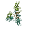
| ||||||||
| Unit cell |
| ||||||||
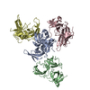
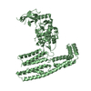
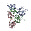
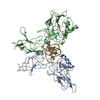
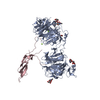
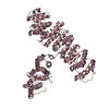
| Details | The biological assembly is a NFAT1-RHR homodimer on DNA. There are three such dimers in the asymmetric unit, related by a local 3 screw axis. DNA stacks continuously in the crystal. |
-Components
| #1: DNA chain | Mass: 4294.814 Da / Num. of mol.: 3 / Source method: obtained synthetically #2: DNA chain | Mass: 4263.804 Da / Num. of mol.: 3 / Source method: obtained synthetically #3: Protein | NFAT / T cell transcription factor NFAT1 / NFAT pre-existing subunit / NF-ATp / RHRMass: 34270.898 Da / Num. of mol.: 6 / Fragment: NFAT1 DNA-binding domain Source method: isolated from a genetically manipulated source Source: (gene. exp.) Homo sapiens (human) / Gene: NFAT1 / Plasmid: PLM1 / Species (production host): Escherichia coli / Production host:  Escherichia coli BL21(DE3) (bacteria) / Strain (production host): BL21 (DE3) / References: UniProt: Q13469 Escherichia coli BL21(DE3) (bacteria) / Strain (production host): BL21 (DE3) / References: UniProt: Q13469 |
|---|
-Experimental details
-Experiment
| Experiment | Method: X-RAY DIFFRACTION / Number of used crystals: 1 |
|---|
- Sample preparation
| Crystal | Density Matthews: 2.61 Å3/Da / Density % sol: 52.88 % | ||||||||||||||||||||||||||||||||||||||||||||||||||||||||||||||||||||||||||||||||||||||||||||||||||||||||||||||||
|---|---|---|---|---|---|---|---|---|---|---|---|---|---|---|---|---|---|---|---|---|---|---|---|---|---|---|---|---|---|---|---|---|---|---|---|---|---|---|---|---|---|---|---|---|---|---|---|---|---|---|---|---|---|---|---|---|---|---|---|---|---|---|---|---|---|---|---|---|---|---|---|---|---|---|---|---|---|---|---|---|---|---|---|---|---|---|---|---|---|---|---|---|---|---|---|---|---|---|---|---|---|---|---|---|---|---|---|---|---|---|---|---|---|
| Crystal grow | Temperature: 292 K / Method: microbatch / pH: 8 Details: HEPES, NaCl, glycerol, NH4OAc, MgCl2, spermine, PEG4000, Tris-HCl, pH 8.0, microbatch, temperature 292.0K | ||||||||||||||||||||||||||||||||||||||||||||||||||||||||||||||||||||||||||||||||||||||||||||||||||||||||||||||||
| Components of the solutions |
| ||||||||||||||||||||||||||||||||||||||||||||||||||||||||||||||||||||||||||||||||||||||||||||||||||||||||||||||||
| Crystal grow | *PLUS pH: 6.3 / Method: vapor diffusion, hanging drop | ||||||||||||||||||||||||||||||||||||||||||||||||||||||||||||||||||||||||||||||||||||||||||||||||||||||||||||||||
| Components of the solutions | *PLUS
|
-Data collection
| Diffraction | Mean temperature: 100 K |
|---|---|
| Diffraction source | Source: SYNCHROTRON / Site: CHESS  / Beamline: F1 / Wavelength: 0.948 Å / Beamline: F1 / Wavelength: 0.948 Å |
| Detector | Type: ADSC QUANTUM 4 / Detector: CCD / Date: Jan 21, 2001 |
| Radiation | Monochromator: Bent triangular asymmetric cut Si(111) monochromater (provides horizontal focussing) Rh-coated Si mirror for vertical focussing Protocol: SINGLE WAVELENGTH / Monochromatic (M) / Laue (L): M / Scattering type: x-ray |
| Radiation wavelength | Wavelength: 0.948 Å / Relative weight: 1 |
| Reflection | Resolution: 3.1→49.11 Å / Num. all: 44978 / Num. obs: 37121 / % possible obs: 82.5 % / Observed criterion σ(F): 0 / Observed criterion σ(I): 0 |
| Reflection shell | Resolution: 3.1→3.29 Å / % possible all: 82.5 |
| Reflection | *PLUS Highest resolution: 3.1 Å / Lowest resolution: 30 Å / Num. obs: 37145 / % possible obs: 82.4 % / Num. measured all: 124267 / Rmerge(I) obs: 0.095 |
| Reflection shell | *PLUS Lowest resolution: 3.2 Å / % possible obs: 77 % / Num. unique obs: 4269 / Num. measured obs: 9841 / Rmerge(I) obs: 0.308 / Mean I/σ(I) obs: 2.7 |
- Processing
| Software |
| ||||||||||||||||||||||||||||||||||||||||||||||||||||||||||||||||||||||||||||||||
|---|---|---|---|---|---|---|---|---|---|---|---|---|---|---|---|---|---|---|---|---|---|---|---|---|---|---|---|---|---|---|---|---|---|---|---|---|---|---|---|---|---|---|---|---|---|---|---|---|---|---|---|---|---|---|---|---|---|---|---|---|---|---|---|---|---|---|---|---|---|---|---|---|---|---|---|---|---|---|---|---|---|
| Refinement | Method to determine structure: MOLECULAR REPLACEMENT / Resolution: 3.1→29.78 Å / Rfactor Rfree error: 0.007 / Occupancy max: 1 / Occupancy min: 0 / Data cutoff high absF: 3392718.05 / Data cutoff high rms absF: 3392718.05 / Data cutoff low absF: 0 / Cross valid method: THROUGHOUT / σ(F): 0 / Stereochemistry target values: Engh & Huber
| ||||||||||||||||||||||||||||||||||||||||||||||||||||||||||||||||||||||||||||||||
| Solvent computation | Solvent model: CNS bulk solvent model used / Bsol: 42.728 Å2 / ksol: 0.266903 e/Å3 | ||||||||||||||||||||||||||||||||||||||||||||||||||||||||||||||||||||||||||||||||
| Displacement parameters | Biso mean: 68.6 Å2
| ||||||||||||||||||||||||||||||||||||||||||||||||||||||||||||||||||||||||||||||||
| Refine Biso | Class: polymer / Treatment: isotropic | ||||||||||||||||||||||||||||||||||||||||||||||||||||||||||||||||||||||||||||||||
| Refine analyze |
| ||||||||||||||||||||||||||||||||||||||||||||||||||||||||||||||||||||||||||||||||
| Refinement step | Cycle: LAST / Resolution: 3.1→29.78 Å
| ||||||||||||||||||||||||||||||||||||||||||||||||||||||||||||||||||||||||||||||||
| Refine LS restraints |
| ||||||||||||||||||||||||||||||||||||||||||||||||||||||||||||||||||||||||||||||||
| LS refinement shell | Resolution: 3.1→3.29 Å / Rfactor Rfree error: 0.022 / Total num. of bins used: 6
| ||||||||||||||||||||||||||||||||||||||||||||||||||||||||||||||||||||||||||||||||
| Xplor file |
| ||||||||||||||||||||||||||||||||||||||||||||||||||||||||||||||||||||||||||||||||
| Refinement | *PLUS Highest resolution: 3.1 Å / Lowest resolution: 30 Å / % reflection Rfree: 5 % | ||||||||||||||||||||||||||||||||||||||||||||||||||||||||||||||||||||||||||||||||
| Solvent computation | *PLUS | ||||||||||||||||||||||||||||||||||||||||||||||||||||||||||||||||||||||||||||||||
| Displacement parameters | *PLUS | ||||||||||||||||||||||||||||||||||||||||||||||||||||||||||||||||||||||||||||||||
| Refine LS restraints | *PLUS
|
