 Movie
Movie Controller

[English] 日本語
 Yorodumi
- PDB-1pz5: Structural basis of peptide-carbohydrate mimicry in an antibody c... -
+ Open data

- Basic information
| Entry | Database: PDB / ID: 1pz5 | ||||||
|---|---|---|---|---|---|---|---|
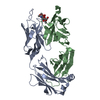
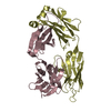
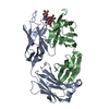
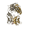
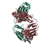
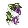
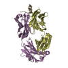
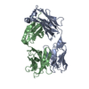
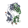
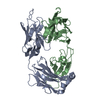
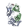
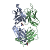
| Title | Structural basis of peptide-carbohydrate mimicry in an antibody combining site | ||||||
 Components Components |
| ||||||
 Keywords Keywords |  IMMUNE SYSTEM / Antibody-antigen structure / peptide-carbohydrate mimicry / vaccine design IMMUNE SYSTEM / Antibody-antigen structure / peptide-carbohydrate mimicry / vaccine design | ||||||
| Function / homology |  Function and homology informationimmunoglobulin complex / immunoglobulin mediated immune response / antigen binding / metal ion binding Function and homology informationimmunoglobulin complex / immunoglobulin mediated immune response / antigen binding / metal ion bindingSimilarity search - Function | ||||||
| Biological species |  Mus musculus (house mouse) Mus musculus (house mouse) | ||||||
| Method | X-RAY DIFFRACTION / SYNCHROTRON / MOLECULAR REPLACEMENT / Resolution: 1.8 Å | ||||||
 Authors Authors | Vyas, N.K. / Vyas, M.N. / Chervenak, M.C. / Bundle, D.R. / Pinto, B.M. / Quiocho, F.A. | ||||||
 Citation Citation | Journal: Proc.Natl.Acad.Sci.USA / Year: 2003 Title: Structural basis of peptide-carbohydrate mimicry in an antibody combining site. Authors: Vyas, N.K. / Vyas, M.N. / Chervenak, M.C. / Bundle, D.R. / Pinto, B.M. / Quiocho, F.A. #1: Journal: Biochemistry / Year: 2002Title: Molecular recognition of oligosaccharide epitopes by a monoclonal Fab sepcific for Shigella flexneri Y lipopolysaccharide: X-ray structures and thermodynamics. Authors: Vyas, N.K. / Vyas, M.N. / Chervenak, M.C. / Johnson, M.A. / Pinto, B.M. / Bundle, D.R. / Quiocho, F.A. #2: Journal: J.Mol.Biol. / Year: 1993Title: Preliminary crystallographic analysis of a Fab specific for the O-antigen of Shigella flexneri cell surface lipopoysaccharide with and without bound saccharides Authors: Vyas, M.N. / Vyas, N.K. / Meikle, P.J. / Sinnott, B. / Pinto, B.M. / Bundle, D.R. / Quiocho, F.A. | ||||||
| History |
| ||||||
| Remark 999 | SEQUENCE No database reference sequence was found for the proteins in this structure at the time of ...SEQUENCE No database reference sequence was found for the proteins in this structure at the time of processing. |
- Structure visualization
| Structure viewer | Molecule: MolmilJmol/JSmol |
|---|
- Downloads & links
-Download
| PDBx/mmCIF format | 1pz5.cif.gz | 109.4 KB | Display | PDBx/mmCIF format |
|---|---|---|---|---|
| PDB format | pdb1pz5.ent.gz | 81.8 KB | Display | PDB format |
| PDBx/mmJSON format | 1pz5.json.gz | Tree view | PDBx/mmJSON format | |
| Others |  Other downloads Other downloads |
-Validation report
| Arichive directory | https://data.pdbj.org/pub/pdb/validation_reports/pz/1pz5ftp://data.pdbj.org/pub/pdb/validation_reports/pz/1pz5 | HTTPS FTP |
|---|
-Related structure data
| Related structure data |  1m71S S: Starting model for refinement |
|---|---|
| Similar structure data |


-Links
 PDBj
PDBj


- Assembly
| Deposited unit | 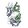
| |||||||||
|---|---|---|---|---|---|---|---|---|---|---|
| 1 |
| |||||||||
| Unit cell |
| |||||||||
| Components on special symmetry positions |
|
-Components
| #1: Antibody | Mass: 23717.350 Da / Num. of mol.: 1 / Source method: isolated from a natural source / Source: (natural) Mus musculus (house mouse) / Organ: spleen / Strain: BALB/C / References: UniProt: A2NHM3*PLUS |
|---|---|
| #2: Antibody | Mass: 23563.404 Da / Num. of mol.: 1 / Source method: isolated from a natural source / Source: (natural) Mus musculus (house mouse) / Organ: spleen / Strain: BALB/C / References: UniProt: P01801*PLUS |
| #3: Protein/peptide | Mass: 976.110 Da / Num. of mol.: 1 / Source method: obtained synthetically Details: The octapeptide was synthesized as its carboxamide (Alberta peptide Institure, Edmonton, Canada) |
| #4: Water | ChemComp-HOH / Water Mass: 18.015 Da / Num. of mol.: 511 / Source method: isolated from a natural source / Formula: H2O Mass: 18.015 Da / Num. of mol.: 511 / Source method: isolated from a natural source / Formula: H2O |
-Experimental details
-Experiment
| Experiment | Method: X-RAY DIFFRACTION / Number of used crystals: 1 |
|---|
- Sample preparation
| Crystal | Density Matthews: 2.47 Å3/Da / Density % sol: 50.19 % |
|---|---|
| Crystal grow | Temperature: 277 K / Method: vapor diffusion / pH: 6.5 Details: MPD, potassium phoshphate, pH 6.5, VAPOR DIFFUSION, temperature 277.0K |
| Crystal grow | *PLUS Details: Vyas, M.N., (1993) J. Mol. Biol., 231, 133. |
-Data collection
| Diffraction source | Source: SYNCHROTRON / Site: NSLS  / Beamline: X4A / Wavelength: 0.948 Å / Beamline: X4A / Wavelength: 0.948 Å |
|---|---|
| Detector | Type: RIGAKU RAXIS IV / Detector: IMAGE PLATE / Details: mirrors |
| Radiation | Protocol: SINGLE WAVELENGTH / Monochromatic (M) / Laue (L): M / Scattering type: x-ray |
| Radiation wavelength | Wavelength: 0.948 Å / Relative weight: 1 |
| Reflection | Resolution: 1.8→20 Å / Num. all: 42148 / Num. obs: 42148 / % possible obs: 91.7 % / Observed criterion σ(F): 0 / Observed criterion σ(I): 0 / Redundancy: 4 % / Biso Wilson estimate: 12.3 Å2 / Rmerge(I) obs: 0.042 / Rsym value: 0.042 / Net I/σ(I): 35.1 |
| Reflection shell | Resolution: 1.8→1.86 Å / Redundancy: 3.65 % / Rmerge(I) obs: 0.12 / Mean I/σ(I) obs: 6.5 / Num. unique all: 2644 / Rsym value: 0.12 / % possible all: 58.9 |
| Reflection | *PLUS Redundancy: 4.02 % / Num. measured all: 169601 |
| Reflection shell | *PLUS % possible obs: 58.9 % / Num. unique obs: 2644 / Num. measured obs: 9660 / Rmerge(I) obs: 0.12 / Mean I/σ(I) obs: 2 |
- Processing
| Software |
| ||||||||||||||||||||||||||||||||||||
|---|---|---|---|---|---|---|---|---|---|---|---|---|---|---|---|---|---|---|---|---|---|---|---|---|---|---|---|---|---|---|---|---|---|---|---|---|---|
| Refinement | Method to determine structure: MOLECULAR REPLACEMENT Starting model: PDB ENTRY 1M71 Resolution: 1.8→19.97 Å / Rfactor Rfree error: 0.004 / Isotropic thermal model: RESTRAINED / Cross valid method: THROUGHOUT / σ(F): 0 / σ(I): 0 / Stereochemistry target values: Engh & Huber
| ||||||||||||||||||||||||||||||||||||
| Solvent computation | Solvent model: FLAT MODEL / Bsol: 48.9066 Å2 / ksol: 0.352158 e/Å3 | ||||||||||||||||||||||||||||||||||||
| Displacement parameters | Biso mean: 20.6 Å2
| ||||||||||||||||||||||||||||||||||||
| Refine analyze | Luzzati coordinate error free: 0.27 Å / Luzzati sigma a free: 0.16 Å | ||||||||||||||||||||||||||||||||||||
| Refinement step | Cycle: LAST / Resolution: 1.8→19.97 Å
| ||||||||||||||||||||||||||||||||||||
| Refine LS restraints |
| ||||||||||||||||||||||||||||||||||||
| LS refinement shell | Resolution: 1.8→1.91 Å / Rfactor Rfree error: 0.012 / Total num. of bins used: 6
| ||||||||||||||||||||||||||||||||||||
| Xplor file |
| ||||||||||||||||||||||||||||||||||||
| Refinement | *PLUS Highest resolution: 1.8 Å / % reflection Rfree: 10 % | ||||||||||||||||||||||||||||||||||||
| Solvent computation | *PLUS | ||||||||||||||||||||||||||||||||||||
| Displacement parameters | *PLUS | ||||||||||||||||||||||||||||||||||||
| Refine LS restraints | *PLUS
| ||||||||||||||||||||||||||||||||||||
| LS refinement shell | *PLUS Highest resolution: 1.8 Å / Rfactor Rwork: 0.233 |
