 Movie
Movie Controller

[English] 日本語
 Yorodumi
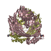
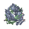
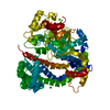
- PDB-1pya: REFINED STRUCTURE OF THE PYRUVOYL-DEPENDENT HISTIDINE DECARBOXYLA... -
+ Open data

- Basic information
| Entry | Database: PDB / ID: 1pya | |||||||||
|---|---|---|---|---|---|---|---|---|---|---|
| Title | REFINED STRUCTURE OF THE PYRUVOYL-DEPENDENT HISTIDINE DECARBOXYLASE FROM LACTOBACILLUS 30A | |||||||||
 Components Components | (PYRUVOYL-DEPENDENT HISTIDINE DECARBOXYLASE (L-HISTIDINE CARBOXYLASE)) x 2 | |||||||||
 Keywords Keywords | CARBOXY-LYASE | |||||||||
| Function / homology |  Function and homology information Function and homology information histidine decarboxylase / histidine decarboxylase activity / histidine metabolic process histidine decarboxylase / histidine decarboxylase activity / histidine metabolic processSimilarity search - Function | |||||||||
| Biological species |  Lactobacillus sp. 30A (bacteria) Lactobacillus sp. 30A (bacteria) | |||||||||
| Method | X-RAY DIFFRACTION / Resolution: 2.5 Å | |||||||||
 Authors Authors | Gallagher, T. / Rozwarski, D.A. / Ernst, S.R. / Hackert, M.L. | |||||||||
 Citation Citation | Journal: J.Mol.Biol. / Year: 1993 Title: Refined structure of the pyruvoyl-dependent histidine decarboxylase from Lactobacillus 30a. Authors: Gallagher, T. / Rozwarski, D.A. / Ernst, S.R. / Hackert, M.L. #1: Journal: J.Biol.Chem. / Year: 1989Title: Pyruvoyl-Dependent Histidine Decarboxylase: Active Site Structure and Mechanistic Analysis Authors: Gallagher, T. / Snell, E.E. / Hackert, M.L. #2: Journal: J.Mol.Biol. / Year: 1985Title: Structure Determination of Histidine Decarboxylase from Lactobacillus 30A at 3.0 Angstroms Resolution Authors: Parks, E.H. / Ernst, S.R. / Hamlin, R. / Xuong, N.H. / Hackert, M.L. #3: Journal: Acta Crystallogr.,Sect.B / Year: 1983Title: The Molecular Symmetry of Histidine Decarboxylase and Prohistidine Decarboxylase by Rotation Function Analysis Authors: Parks, E.H. / Clinger, K. / Hackert, M.L. #4: Journal: J.Biol.Chem. / Year: 1981Title: Crystallization and Subunit Structure of Histidine Decarboxylase from Lactobacillus 30A Authors: Hackert, M.L. / Meador, W.E. / Oliver, R.M. / Salmon, J.B. / Rescei, P.A. / Snell, E.E. | |||||||||
| History |
| |||||||||
| Remark 650 | HELIX ISOLATED TURNS OF A 3-10 HELIX OCCUR AT RESIDUES 98 - 102 AND 266 - 270. |
- Structure visualization
| Structure viewer | Molecule: MolmilJmol/JSmol |
|---|
- Downloads & links
-Download
| PDBx/mmCIF format | 1pya.cif.gz | 246 KB | Display | PDBx/mmCIF format |
|---|---|---|---|---|
| PDB format | pdb1pya.ent.gz | 200.6 KB | Display | PDB format |
| PDBx/mmJSON format | 1pya.json.gz | Tree view | PDBx/mmJSON format | |
| Others |  Other downloads Other downloads |
-Validation report
| Arichive directory | https://data.pdbj.org/pub/pdb/validation_reports/py/1pyaftp://data.pdbj.org/pub/pdb/validation_reports/py/1pya | HTTPS FTP |
|---|
-Related structure data
| Similar structure data |
|---|
-Links
 PDBj
PDBj- Assembly
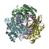
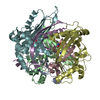
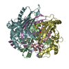
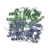
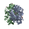
| Deposited unit | 
| ||||||||||||
|---|---|---|---|---|---|---|---|---|---|---|---|---|---|
| 1 |
| ||||||||||||
| Unit cell |
| ||||||||||||
| Noncrystallographic symmetry (NCS) | NCS oper:
| ||||||||||||
| Details | TWO TRIMERS RELATED BY A CRYSTALLOGRAPHIC TWO-FOLD SYMMETRY AXIS GENERATE THE HEXAMER. THE TRANSFORMATION PRESENTED AS *MTRIX 1* BELOW WILL YIELD APPROXIMATE COORDINATES FOR CHAINS *A* AND *B* WHEN APPLIED TO CHAINS *C* AND *D*, RESPECTIVELY. THE TRANSFORMATION PRESENTED AS *MTRIX 2* BELOW WILL YIELD APPROXIMATE COORDINATES FOR CHAINS *A* AND *B* WHEN APPLIED TO CHAINS *E* AND *F*, RESPECTIVELY. |
-Components
| #1: Protein | Mass: 8850.832 Da / Num. of mol.: 3 Source method: isolated from a genetically manipulated source Source: (gene. exp.) Lactobacillus sp. 30A (bacteria) / Strain: 30a / References: UniProt: P00862, histidine decarboxylase#2: Protein | Mass: 25285.375 Da / Num. of mol.: 3 Source method: isolated from a genetically manipulated source Source: (gene. exp.) Lactobacillus sp. 30A (bacteria) / Strain: 30a / References: UniProt: P00862, histidine decarboxylase#3: Water | ChemComp-HOH / | Water Mass: 18.015 Da / Num. of mol.: 471 / Source method: isolated from a natural source / Formula: H2O Mass: 18.015 Da / Num. of mol.: 471 / Source method: isolated from a natural source / Formula: H2O |
|---|
-Experimental details
-Experiment
| Experiment | Method: X-RAY DIFFRACTION |
|---|
- Sample preparation
| Crystal | Density Matthews: 3.21 Å3/Da / Density % sol: 61.71 % | |||||||||||||||||||||||||
|---|---|---|---|---|---|---|---|---|---|---|---|---|---|---|---|---|---|---|---|---|---|---|---|---|---|---|
| Crystal grow | *PLUS pH: 4.8 / Method: unknown | |||||||||||||||||||||||||
| Components of the solutions | *PLUS
|
-Data collection
| Radiation | Scattering type: x-ray |
|---|---|
| Radiation wavelength | Relative weight: 1 |
- Processing
| Software |
| ||||||||||||||||||||||||||||||||||||||||||||||||||||||||||||
|---|---|---|---|---|---|---|---|---|---|---|---|---|---|---|---|---|---|---|---|---|---|---|---|---|---|---|---|---|---|---|---|---|---|---|---|---|---|---|---|---|---|---|---|---|---|---|---|---|---|---|---|---|---|---|---|---|---|---|---|---|---|
| Refinement | Rfactor Rwork: 0.15 / Rfactor obs: 0.15 / Highest resolution: 2.5 Å Details: THE SEGMENT NUMBERS ARE AS FOLLOWS: (1,3,5 = BETA SUBUNITS) (2,4,6 = ALPHA SUBUNITS), (7,8,9 = PVL MOIETIES WITH 7-2 REPRESENTING THE PVL GROUP COVALENTLY ATTACHED TO THE BEGINNING OF AN ...Details: THE SEGMENT NUMBERS ARE AS FOLLOWS: (1,3,5 = BETA SUBUNITS) (2,4,6 = ALPHA SUBUNITS), (7,8,9 = PVL MOIETIES WITH 7-2 REPRESENTING THE PVL GROUP COVALENTLY ATTACHED TO THE BEGINNING OF AN ALPHA SUBUNIT OR SEGMENT 2). THE SEGMENT NUMBERS FOR THE THREE *AB* SUBUNITS ARE: 1, 7 - 2; 3, 8 - 4; AND 5, 9 - 6. | ||||||||||||||||||||||||||||||||||||||||||||||||||||||||||||
| Refinement step | Cycle: LAST / Highest resolution: 2.5 Å
| ||||||||||||||||||||||||||||||||||||||||||||||||||||||||||||
| Refine LS restraints |
| ||||||||||||||||||||||||||||||||||||||||||||||||||||||||||||
| Software | *PLUS Name: X-PLOR / Classification: refinement | ||||||||||||||||||||||||||||||||||||||||||||||||||||||||||||
| Refinement | *PLUS Highest resolution: 2.5 Å / Lowest resolution: 5 Å / Num. reflection obs: 39926 / Rfactor obs: 0.15 | ||||||||||||||||||||||||||||||||||||||||||||||||||||||||||||
| Solvent computation | *PLUS | ||||||||||||||||||||||||||||||||||||||||||||||||||||||||||||
| Displacement parameters | *PLUS | ||||||||||||||||||||||||||||||||||||||||||||||||||||||||||||
| Refine LS restraints | *PLUS
|
