 Movie
Movie Controller


+ Open data

- Basic information
| Entry | Database: PDB / ID: 1psw | ||||||
|---|---|---|---|---|---|---|---|
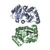
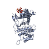
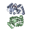
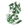
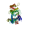
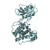
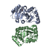
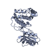
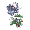
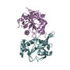
| Title | Structure of E. coli ADP-heptose lps heptosyltransferase II | ||||||
 Components Components | ADP-HEPTOSE LPS HEPTOSYLTRANSFERASE II | ||||||
 Keywords Keywords |  TRANSFERASE / STRUCTURAL GENOMICS / T832 / NYSGXRC / LPS BIOSYNTHETIC PATHWAY / PSI / Protein Structure Initiative / New York SGX Research Center for Structural Genomics TRANSFERASE / STRUCTURAL GENOMICS / T832 / NYSGXRC / LPS BIOSYNTHETIC PATHWAY / PSI / Protein Structure Initiative / New York SGX Research Center for Structural Genomics | ||||||
| Function / homology |  Function and homology information Function and homology informationADP-heptose-lipopolysaccharide heptosyltransferase activity / Transferases / lipopolysaccharide core region biosynthetic process / cytosolSimilarity search - Function | ||||||
| Biological species |  Escherichia coli (E. coli) Escherichia coli (E. coli) | ||||||
| Method | X-RAY DIFFRACTION / SYNCHROTRON / SAD / Resolution: 2 Å | ||||||
 Authors Authors | Kniewel, R. / Buglino, J. / Solorzano, V. / Wu, J. / Lima, C.D. / Burley, S.K. / New York SGX Research Center for Structural Genomics (NYSGXRC) | ||||||
 Citation Citation | Journal: To be Published Title: Structure of E. coli ADP-heptose lps heptosyltransferase II Authors: Kniewel, R. / Buglino, J. / Solorzano, V. / Wu, J. / Lima, C.D. | ||||||
| History |
|
- Structure visualization
| Structure viewer | Molecule: MolmilJmol/JSmol |
|---|
- Downloads & links
-Download
| PDBx/mmCIF format | 1psw.cif.gz | 81.1 KB | Display | PDBx/mmCIF format |
|---|---|---|---|---|
| PDB format | pdb1psw.ent.gz | 64.4 KB | Display | PDB format |
| PDBx/mmJSON format | 1psw.json.gz | Tree view | PDBx/mmJSON format | |
| Others |  Other downloads Other downloads |
-Validation report
| Arichive directory | https://data.pdbj.org/pub/pdb/validation_reports/ps/1pswftp://data.pdbj.org/pub/pdb/validation_reports/ps/1psw | HTTPS FTP |
|---|
-Related structure data
| Similar structure data | |
|---|---|
| Other databases |
-Links
 PDBj
PDBj- Assembly
| Deposited unit | 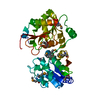
| ||||||||||
|---|---|---|---|---|---|---|---|---|---|---|---|
| 1 |
| ||||||||||
| Unit cell |
|
-Components
| #1: Protein | Mass: 39617.785 Da / Num. of mol.: 1 Source method: isolated from a genetically manipulated source Source: (gene. exp.) Escherichia coli (E. coli) / Gene: rfaF / Plasmid: PET T7 / Production host: Escherichia coli (E. coli) / Strain (production host): B834 DE3 / References: UniProt: P37692, EC: 2.4.99.- |
|---|---|
| #2: Water | ChemComp-HOH / Water Mass: 18.015 Da / Num. of mol.: 283 / Source method: isolated from a natural source / Formula: H2O Mass: 18.015 Da / Num. of mol.: 283 / Source method: isolated from a natural source / Formula: H2O |
-Experimental details
-Experiment
| Experiment | Method: X-RAY DIFFRACTION / Number of used crystals: 1 |
|---|
- Sample preparation
| Crystal | Density Matthews: 2.91 Å3/Da / Density % sol: 57.4 % |
|---|---|
| Crystal grow | Temperature: 291 K / Method: vapor diffusion / pH: 5.6 Details: 1M Ammonium Phospate, 0.1M, Na Citrate pH 5.6 , VAPOR DIFFUSION, temperature 291K |
-Data collection
| Diffraction | Mean temperature: 100 K |
|---|---|
| Diffraction source | Source: SYNCHROTRON / Site: APS  / Beamline: 31-ID / Wavelength: 0.9798 Å / Beamline: 31-ID / Wavelength: 0.9798 Å |
| Detector | Type: MARRESEARCH / Detector: CCD / Date: Jun 7, 2003 |
| Radiation | Monochromator: SAGITALLY FOCUSED Si(111) / Protocol: SAD / Monochromatic (M) / Laue (L): M / Scattering type: x-ray |
| Radiation wavelength | Wavelength: 0.9798 Å / Relative weight: 1 |
| Reflection | Resolution: 1.9→20 Å / Num. all: 37604 / Num. obs: 37529 / % possible obs: 99.8 % / Observed criterion σ(I): -3 / Biso Wilson estimate: 24.6 Å2 / Rmerge(I) obs: 0.093 |
| Reflection shell | Resolution: 1.9→1.97 Å / % possible all: 100 |
- Processing
| Software |
| ||||||||||||||||||||||||||||||||||||
|---|---|---|---|---|---|---|---|---|---|---|---|---|---|---|---|---|---|---|---|---|---|---|---|---|---|---|---|---|---|---|---|---|---|---|---|---|---|
| Refinement | Method to determine structure: SAD / Resolution: 2→19.96 Å / Rfactor Rfree error: 0.006 / Isotropic thermal model: RESTRAINED / Cross valid method: THROUGHOUT / σ(F): 0 / Stereochemistry target values: Engh & Huber / Details: BULK SOLVENT MODEL USED
| ||||||||||||||||||||||||||||||||||||
| Solvent computation | Solvent model: FLAT MODEL / Bsol: 52.3843 Å2 / ksol: 0.381507 e/Å3 | ||||||||||||||||||||||||||||||||||||
| Displacement parameters | Biso mean: 29 Å2
| ||||||||||||||||||||||||||||||||||||
| Refine analyze | Luzzati coordinate error free: 0.24 Å / Luzzati sigma a free: 0.14 Å | ||||||||||||||||||||||||||||||||||||
| Refinement step | Cycle: LAST / Resolution: 2→19.96 Å
| ||||||||||||||||||||||||||||||||||||
| Refine LS restraints |
| ||||||||||||||||||||||||||||||||||||
| LS refinement shell | Resolution: 2→2.13 Å / Rfactor Rfree error: 0.016 / Total num. of bins used: 6
| ||||||||||||||||||||||||||||||||||||
| Xplor file |
|
