 Movie
Movie Controller


+ Open data

- Basic information
| Entry | Database: PDB / ID: 1pre | ||||||
|---|---|---|---|---|---|---|---|
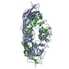
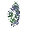
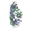
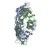
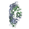
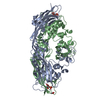
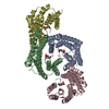
| Title | PROAEROLYSIN | ||||||
 Components Components | PROAEROLYSIN | ||||||
 Keywords Keywords | TOXIN (HEMOLYTIC POLYPEPTIDE) | ||||||
| Function / homology |  Function and homology information Function and homology information toxin activity / host cell plasma membrane / extracellular region / membrane / identical protein binding toxin activity / host cell plasma membrane / extracellular region / membrane / identical protein bindingSimilarity search - Function | ||||||
| Biological species |  Aeromonas hydrophila (bacteria) Aeromonas hydrophila (bacteria) | ||||||
| Method | X-RAY DIFFRACTION / SYNCHROTRON / Resolution: 2.8 Å | ||||||
 Authors Authors | Parker, M.W. / Buckley, J.T. / Postma, J.P.M. / Tucker, A.D. / Tsernoglou, D. | ||||||
 Citation Citation | Journal: Nature / Year: 1994 Title: Structure of the Aeromonas toxin proaerolysin in its water-soluble and membrane-channel states. Authors: Parker, M.W. / Buckley, J.T. / Postma, J.P. / Tucker, A.D. / Leonard, K. / Pattus, F. / Tsernoglou, D. #1: Journal: To be PublishedTitle: Structural Analysis of Proaerolysin and Various Single-Site Mutants as a Basis for Understanding Membrane Insertion of the Toxin Authors: Parker, M.W. / Buckley, J.T. / Postma, J.P.M. / Feil, S.C. / Van Der Goot, F.G. / Vetter, I. / Tucker, A.D. / Tsernoglou, D. #2: Journal: J.Mol.Biol. / Year: 1990Title: Crystallization of a Proform of Aerolysin, a Hole-Forming Toxin from Aeromonas Hydrophila Authors: Tucker, A.D. / Parker, M.W. / Tsernoglou, D. / Buckley, J.T. | ||||||
| History |
|
- Structure visualization
| Structure viewer | Molecule: MolmilJmol/JSmol |
|---|
- Downloads & links
-Download
| PDBx/mmCIF format | 1pre.cif.gz | 176.5 KB | Display | PDBx/mmCIF format |
|---|---|---|---|---|
| PDB format | pdb1pre.ent.gz | 147 KB | Display | PDB format |
| PDBx/mmJSON format | 1pre.json.gz | Tree view | PDBx/mmJSON format | |
| Others |  Other downloads Other downloads |
-Validation report
| Arichive directory | https://data.pdbj.org/pub/pdb/validation_reports/pr/1preftp://data.pdbj.org/pub/pdb/validation_reports/pr/1pre | HTTPS FTP |
|---|
-Related structure data
| Similar structure data |
|---|

-Links
 PDBj
PDBj


- Assembly
| Deposited unit | 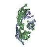
| ||||||||
|---|---|---|---|---|---|---|---|---|---|
| 1 |
| ||||||||
| Unit cell |
| ||||||||
| Noncrystallographic symmetry (NCS) | NCS oper: (Code: given Matrix: (-1, -0.00043, 0.00286), Vector : |
-Components
| #1: Protein | Mass: 51980.453 Da / Num. of mol.: 2 Source method: isolated from a genetically manipulated source Source: (gene. exp.) Aeromonas hydrophila (bacteria) / Plasmid: PRK2013 / Production host: Aeromonas salmonicida (bacteria) / References: UniProt: P09167#2: Water | ChemComp-HOH / | Water Mass: 18.015 Da / Num. of mol.: 33 / Source method: isolated from a natural source / Formula: H2O Mass: 18.015 Da / Num. of mol.: 33 / Source method: isolated from a natural source / Formula: H2O |
|---|
-Experimental details
-Experiment
| Experiment | Method: X-RAY DIFFRACTION |
|---|
- Sample preparation
| Crystal | Density Matthews: 2.89 Å3/Da / Density % sol: 57 % | ||||||||||||||||||||||||
|---|---|---|---|---|---|---|---|---|---|---|---|---|---|---|---|---|---|---|---|---|---|---|---|---|---|
| Crystal grow | pH: 5.6 / Details: pH 5.6 | ||||||||||||||||||||||||
| Crystal | *PLUS | ||||||||||||||||||||||||
| Crystal grow | *PLUS Method: vapor diffusion, hanging drop / Details: Tucker, A.D., (1990) J.Mol.Biol., 212, 561. / pH: 5.4 | ||||||||||||||||||||||||
| Components of the solutions | *PLUS
|
-Data collection
| Diffraction source | Source: SYNCHROTRON / Site: EMBL/DESY, Hamburg  / Beamline: X11 / Wavelength: 1.009 / Beamline: X11 / Wavelength: 1.009 |
|---|---|
| Detector | Type: MARRESEARCH / Detector: IMAGE PLATE / Date: Nov 15, 1989 |
| Radiation | Monochromatic (M) / Laue (L): M / Scattering type: x-ray |
| Radiation wavelength | Wavelength: 1.009 Å / Relative weight: 1 |
| Reflection | Resolution: 2.8→40 Å / Num. obs: 30060 / % possible obs: 98 % / Observed criterion σ(I): 0 / Redundancy: 4.9 % / Rmerge(I) obs: 0.091 |
| Reflection | *PLUS Num. measured all: 145443 |
- Processing
| Software |
| ||||||||||||||||||||||||||||||||||||||||||||||||||||||||||||||||||||||||||||||||||||
|---|---|---|---|---|---|---|---|---|---|---|---|---|---|---|---|---|---|---|---|---|---|---|---|---|---|---|---|---|---|---|---|---|---|---|---|---|---|---|---|---|---|---|---|---|---|---|---|---|---|---|---|---|---|---|---|---|---|---|---|---|---|---|---|---|---|---|---|---|---|---|---|---|---|---|---|---|---|---|---|---|---|---|---|---|---|
| Refinement | Resolution: 2.8→6 Å / σ(F): 0 /
| ||||||||||||||||||||||||||||||||||||||||||||||||||||||||||||||||||||||||||||||||||||
| Displacement parameters | Biso mean: 33.8 Å2 | ||||||||||||||||||||||||||||||||||||||||||||||||||||||||||||||||||||||||||||||||||||
| Refinement step | Cycle: LAST / Resolution: 2.8→6 Å
| ||||||||||||||||||||||||||||||||||||||||||||||||||||||||||||||||||||||||||||||||||||
| Refine LS restraints |
| ||||||||||||||||||||||||||||||||||||||||||||||||||||||||||||||||||||||||||||||||||||
| Software | *PLUS Name: PROLSQ / Classification: refinement | ||||||||||||||||||||||||||||||||||||||||||||||||||||||||||||||||||||||||||||||||||||
| Refinement | *PLUS Rfactor obs: 0.208 | ||||||||||||||||||||||||||||||||||||||||||||||||||||||||||||||||||||||||||||||||||||
| Solvent computation | *PLUS | ||||||||||||||||||||||||||||||||||||||||||||||||||||||||||||||||||||||||||||||||||||
| Displacement parameters | *PLUS |
