 Movie
Movie Controller

[English] 日本語
 Yorodumi
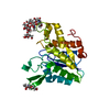
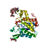
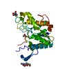
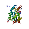
- PDB-1pp4: The crystal structure of rhamnogalacturonan acetylesterase in spa... -
+ Open data

- Basic information
| Entry | Database: PDB / ID: 1pp4 | ||||||
|---|---|---|---|---|---|---|---|
| Title | The crystal structure of rhamnogalacturonan acetylesterase in space group P3121 | ||||||
 Components Components | Rhamnogalacturonan acetylesterase | ||||||
 Keywords Keywords | HYDROLASE / GDS(L) hydrolase | ||||||
| Function / homology |  Function and homology informationrhamnogalacturonan acetylesterase / hydrolase activity, acting on ester bonds Function and homology informationrhamnogalacturonan acetylesterase / hydrolase activity, acting on ester bondsSimilarity search - Function | ||||||
| Biological species |  Aspergillus aculeatus (mold) Aspergillus aculeatus (mold) | ||||||
| Method | X-RAY DIFFRACTION / MOLECULAR REPLACEMENT / Resolution: 2.5 Å | ||||||
 Authors Authors | Molgaard, A. / Larsen, S. | ||||||
 Citation Citation | Journal: Acta Crystallogr.,Sect.D / Year: 2004 Title: Crystal packing in two pH-dependent crystal forms of rhamnogalacturonan acetylesterase. Authors: Molgaard, A. / Larsen, S. #1: Journal: Structure Fold.Des. / Year: 2000Title: Rhamnogalacturonan acetylesterase elucidates the structure and function of a new family of hydrolases Authors: Molgaard, A. / Kauppinen, S. / Larsen, S. #2: Journal: Acta Crystallogr.,Sect.D / Year: 1998Title: Crystallization and preliminary X-ray diffraction studies of the heterogeneously glycosylated enzyme rhamnogalacturonan acetylesterase from Aspergillus aculeatus Authors: Molgaard, A. / Petersen, J.F. / Kauppinen, S. / Dalboge, H. / Johnsen, A.H. / Poulsen, J.C.N. / Larsen, S. #3: Journal: Acta Crystallogr.,Sect.D / Year: 2002Title: A branched N-linked glycan at atomic resolution in the 1.12 A structure of rhamnogalacturonan acetylesterase Authors: Molgaard, A. / Larsen, S. #4: Journal: J.Biol.Chem. / Year: 1995Title: Molecular cloning and characterization of a rhamnogalacturonan acetylesterase from Aspergillus aculeatus. Synergism between rhamnogalacturonan degrading enzymes Authors: Kauppinen, S. / Christgau, S. / Kofod, L.V. / Halkier, T. / Dorreich, K. / Dalboge, H. | ||||||
| History |
|
- Structure visualization
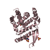
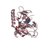
| Structure viewer | Molecule: MolmilJmol/JSmol |
|---|
- Downloads & links
-Download
| PDBx/mmCIF format | 1pp4.cif.gz | 99.4 KB | Display | PDBx/mmCIF format |
|---|---|---|---|---|
| PDB format | pdb1pp4.ent.gz | 76.6 KB | Display | PDB format |
| PDBx/mmJSON format | 1pp4.json.gz | Tree view | PDBx/mmJSON format | |
| Others |  Other downloads Other downloads |
-Validation report
| Arichive directory | https://data.pdbj.org/pub/pdb/validation_reports/pp/1pp4ftp://data.pdbj.org/pub/pdb/validation_reports/pp/1pp4 | HTTPS FTP |
|---|
-Related structure data
| Related structure data | 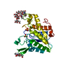 1deoS S: Starting model for refinement |
|---|---|
| Similar structure data |


-Links
 PDBj
PDBj- Assembly
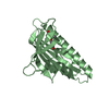
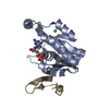
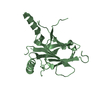
| Deposited unit | 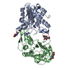
| ||||||||
|---|---|---|---|---|---|---|---|---|---|
| 1 | 
| ||||||||
| 2 | 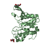
| ||||||||
| Unit cell |
| ||||||||
| Details | The biological assembly is a monomer represented by either the A or the B chain in the asymmetric unit |
-Components
| #1: Protein | / RGAE Mass: 24622.881 Da / Num. of mol.: 2 Source method: isolated from a genetically manipulated source Source: (gene. exp.) Aspergillus aculeatus (mold) / Gene: RHA1 / Plasmid: PHD464 / Production host: Aspergillus oryzae (mold) / Strain (production host): KSM 510References: UniProt: Q00017, Hydrolases; Acting on ester bonds; Carboxylic-ester hydrolases#2: Sugar | ChemComp-NAG / N-Acetylglucosamine  Type: D-saccharide, beta linking / Mass: 221.208 Da / Num. of mol.: 4 Type: D-saccharide, beta linking / Mass: 221.208 Da / Num. of mol.: 4Source method: isolated from a genetically manipulated source Formula: C8H15NO6 #3: Water | ChemComp-HOH / | Water Mass: 18.015 Da / Num. of mol.: 59 / Source method: isolated from a natural source / Formula: H2O Mass: 18.015 Da / Num. of mol.: 59 / Source method: isolated from a natural source / Formula: H2O |
|---|
-Experimental details
-Experiment
| Experiment | Method: X-RAY DIFFRACTION / Number of used crystals: 1 |
|---|
- Sample preparation
| Crystal | Density Matthews: 3.53 Å3/Da / Density % sol: 65.18 % |
|---|---|
| Crystal grow | Temperature: 298 K / Method: vapor diffusion, hanging drop / pH: 4.5 Details: PEG 4000, 2-propanol, citrate, pH 4.5, VAPOR DIFFUSION, HANGING DROP, temperature 298K |
-Data collection
| Diffraction | Mean temperature: 291 K |
|---|---|
| Diffraction source | Source: ROTATING ANODE / Type: RIGAKU / Wavelength: 1.5418 Å |
| Detector | Type: RIGAKU RAXIS II / Detector: IMAGE PLATE / Date: Jul 15, 1996 |
| Radiation | Monochromator: graphite / Protocol: SINGLE WAVELENGTH / Monochromatic (M) / Laue (L): M / Scattering type: x-ray |
| Radiation wavelength | Wavelength: 1.5418 Å / Relative weight: 1 |
| Reflection | Resolution: 2.5→29.6 Å / Num. all: 25036 / Num. obs: 24979 / % possible obs: 98.8 % / Observed criterion σ(F): 0 / Observed criterion σ(I): 0 / Redundancy: 5 % / Biso Wilson estimate: 56.79 Å2 / Rmerge(I) obs: 0.087 |
| Reflection shell | Resolution: 2.49→2.62 Å / Redundancy: 5.6 % / Rmerge(I) obs: 0.452 / % possible all: 93 |
- Processing
| Software |
| ||||||||||||||||||||
|---|---|---|---|---|---|---|---|---|---|---|---|---|---|---|---|---|---|---|---|---|---|
| Refinement | Method to determine structure: MOLECULAR REPLACEMENT Starting model: pdb entry 1deo Resolution: 2.5→28.18 Å / σ(F): 2 / Stereochemistry target values: Engh & Huber
| ||||||||||||||||||||
| Refinement step | Cycle: LAST / Resolution: 2.5→28.18 Å
| ||||||||||||||||||||
| Refine LS restraints |
| ||||||||||||||||||||
| LS refinement shell | Resolution: 2.5→2.61 Å /
|
