 Movie
Movie Controller

[English] 日本語
 Yorodumi
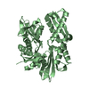
- PDB-1pm4: Crystal structure of Yersinia pseudotuberculosis-derived mitogen (YPM) -
+ Open data

- Basic information
| Entry | Database: PDB / ID: 1pm4 | ||||||
|---|---|---|---|---|---|---|---|
| Title | Crystal structure of Yersinia pseudotuberculosis-derived mitogen (YPM) | ||||||
 Components Components | YPM | ||||||
 Keywords Keywords |  TOXIN / Jelly roll fold TOXIN / Jelly roll fold | ||||||
| Function / homology | Mitogen Ypm / Mitogen, Yersinia pseudotuberculosis / Mitogen Ypm superfamily / Yersinia pseudo-tuberculosis mitogen / Jelly Rolls / Sandwich / Mainly Beta / -derived mitogen Function and homology information Function and homology information | ||||||
| Biological species |  Yersinia pseudotuberculosis (bacteria) Yersinia pseudotuberculosis (bacteria) | ||||||
| Method | X-RAY DIFFRACTION / SYNCHROTRON / MAD / Resolution: 1.755 Å | ||||||
 Authors Authors | Donadini, R. / Liew, C.W. / Kwan, A.H. / Mackay, J.P. / Fields, B.A. | ||||||
 Citation Citation | Journal: Structure / Year: 2004 Title: Crystal and Solution Structures of a Superantigen from Yersinia pseudotuberculosis Reveal a Jelly-Roll Fold. Authors: Donadini, R. / Liew, C.W. / Kwan, A.H. / Mackay, J.P. / Fields, B.A. | ||||||
| History |
| ||||||
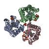
| Remark 300 | BIOMOLECULE: 1 THIS ENTRY CONTAINS THE CRYSTALLOGRAPHIC ASYMMETRIC UNIT WHICH CONSISTS OF 3 ... BIOMOLECULE: 1 THIS ENTRY CONTAINS THE CRYSTALLOGRAPHIC ASYMMETRIC UNIT WHICH CONSISTS OF 3 CHAIN(S). The trimerization may be biologically relevant but has not yet been established experimentally. The protein is a monomer in solution. |
- Structure visualization
| Structure viewer | Molecule: MolmilJmol/JSmol |
|---|
- Downloads & links
-Download
| PDBx/mmCIF format | 1pm4.cif.gz | 83.2 KB | Display | PDBx/mmCIF format |
|---|---|---|---|---|
| PDB format | pdb1pm4.ent.gz | 67.2 KB | Display | PDB format |
| PDBx/mmJSON format | 1pm4.json.gz | Tree view | PDBx/mmJSON format | |
| Others |  Other downloads Other downloads |
-Validation report
| Arichive directory | https://data.pdbj.org/pub/pdb/validation_reports/pm/1pm4ftp://data.pdbj.org/pub/pdb/validation_reports/pm/1pm4 | HTTPS FTP |
|---|
-Related structure data


-Links
 PDBj
PDBj- Assembly
| Deposited unit | 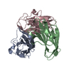
| ||||||||
|---|---|---|---|---|---|---|---|---|---|
| 1 |
| ||||||||
| Unit cell |
|
-Components
| #1: Protein | Mass: 13223.785 Da / Num. of mol.: 3 Source method: isolated from a genetically manipulated source Source: (gene. exp.) Yersinia pseudotuberculosis (bacteria) / Gene: ypma / Plasmid: pMAL-p2x / Production host: Escherichia coli (E. coli) / Strain (production host): Novablue / References: UniProt: Q57221#2: Water | ChemComp-HOH / | Water Mass: 18.015 Da / Num. of mol.: 339 / Source method: isolated from a natural source / Formula: H2O Mass: 18.015 Da / Num. of mol.: 339 / Source method: isolated from a natural source / Formula: H2O |
|---|
-Experimental details
-Experiment
| Experiment | Method: X-RAY DIFFRACTION / Number of used crystals: 1 |
|---|
- Sample preparation
| Crystal | Density Matthews: 2.26 Å3/Da / Density % sol: 45.55 % | ||||||||||||||||||||||||||||||||||||
|---|---|---|---|---|---|---|---|---|---|---|---|---|---|---|---|---|---|---|---|---|---|---|---|---|---|---|---|---|---|---|---|---|---|---|---|---|---|
| Crystal grow | Temperature: 293 K / Method: vapor diffusion, sitting drop / pH: 5.8 Details: Ammonium sulfate, PEG 200, sodium citrate, pH 5.8, VAPOR DIFFUSION, SITTING DROP, temperature 293K | ||||||||||||||||||||||||||||||||||||
| Crystal grow | *PLUS Temperature: 293 K / pH: 7.2 / Method: vapor diffusion, sitting drop / Details: Donadini, R., (2003) Acta Cryst., D59, 1330. | ||||||||||||||||||||||||||||||||||||
| Components of the solutions | *PLUS
|
-Data collection
| Diffraction | Mean temperature: 100 K |
|---|---|
| Diffraction source | Source: SYNCHROTRON / Site: SSRL  / Beamline: BL1-5 / Wavelength: 1.071 Å / Beamline: BL1-5 / Wavelength: 1.071 Å |
| Detector | Type: ADSC QUANTUM 4 / Detector: CCD / Date: Apr 12, 2002 |
| Radiation | Monochromator: 2-crystal monochromator, Si111, 1m long Rh coated bent cylindrical mirror for horizontal and vertical focussing Protocol: SINGLE WAVELENGTH / Monochromatic (M) / Laue (L): M / Scattering type: x-ray |
| Radiation wavelength | Wavelength: 1.071 Å / Relative weight: 1 |
| Reflection | Resolution: 1.75→40 Å / Num. all: 253851 / Num. obs: 253092 / % possible obs: 99.7 % / Observed criterion σ(F): 0 / Observed criterion σ(I): 0 / Redundancy: 7.2 % / Rmerge(I) obs: 0.045 / Net I/σ(I): 39.1 |
| Reflection shell | Resolution: 1.755→1.8 Å / Rmerge(I) obs: 0.515 / Mean I/σ(I) obs: 3.2 / % possible all: 99.1 |
| Reflection | *PLUS Num. obs: 35166 / Num. measured all: 253092 |
| Reflection shell | *PLUS Highest resolution: 1.75 Å / % possible obs: 99.1 % |
- Processing
| Software |
| ||||||||||||||||||||||||||||||||||||||||||||||||||||||||||||||||||||||||||||||||||||||||||||||||||||
|---|---|---|---|---|---|---|---|---|---|---|---|---|---|---|---|---|---|---|---|---|---|---|---|---|---|---|---|---|---|---|---|---|---|---|---|---|---|---|---|---|---|---|---|---|---|---|---|---|---|---|---|---|---|---|---|---|---|---|---|---|---|---|---|---|---|---|---|---|---|---|---|---|---|---|---|---|---|---|---|---|---|---|---|---|---|---|---|---|---|---|---|---|---|---|---|---|---|---|---|---|---|
| Refinement | Method to determine structure: MAD / Resolution: 1.755→34.71 Å / Cor.coef. Fo:Fc: 0.958 / Cor.coef. Fo:Fc free: 0.93 / SU B: 2.441 / SU ML: 0.079 / Cross valid method: THROUGHOUT / σ(F): 0 / ESU R: 0.125 / ESU R Free: 0.125 / Details: HYDROGENS HAVE BEEN ADDED IN THE RIDING POSITIONS
| ||||||||||||||||||||||||||||||||||||||||||||||||||||||||||||||||||||||||||||||||||||||||||||||||||||
| Solvent computation | Ion probe radii: 0.8 Å / Shrinkage radii: 0.8 Å / VDW probe radii: 1.4 Å / Solvent model: BABINET MODEL WITH MASK | ||||||||||||||||||||||||||||||||||||||||||||||||||||||||||||||||||||||||||||||||||||||||||||||||||||
| Displacement parameters | Biso mean: 18.498 Å2
| ||||||||||||||||||||||||||||||||||||||||||||||||||||||||||||||||||||||||||||||||||||||||||||||||||||
| Refinement step | Cycle: LAST / Resolution: 1.755→34.71 Å
| ||||||||||||||||||||||||||||||||||||||||||||||||||||||||||||||||||||||||||||||||||||||||||||||||||||
| Refine LS restraints |
| ||||||||||||||||||||||||||||||||||||||||||||||||||||||||||||||||||||||||||||||||||||||||||||||||||||
| LS refinement shell | Resolution: 1.755→1.8 Å / Total num. of bins used: 20 /
| ||||||||||||||||||||||||||||||||||||||||||||||||||||||||||||||||||||||||||||||||||||||||||||||||||||
| Refinement | *PLUS Highest resolution: 1.75 Å / Lowest resolution: 40 Å / Rfactor Rfree: 0.229 / Rfactor Rwork: 0.182 | ||||||||||||||||||||||||||||||||||||||||||||||||||||||||||||||||||||||||||||||||||||||||||||||||||||
| Solvent computation | *PLUS | ||||||||||||||||||||||||||||||||||||||||||||||||||||||||||||||||||||||||||||||||||||||||||||||||||||
| Displacement parameters | *PLUS | ||||||||||||||||||||||||||||||||||||||||||||||||||||||||||||||||||||||||||||||||||||||||||||||||||||
| Refine LS restraints | *PLUS
| ||||||||||||||||||||||||||||||||||||||||||||||||||||||||||||||||||||||||||||||||||||||||||||||||||||
| LS refinement shell | *PLUS Highest resolution: 1.75 Å / Lowest resolution: 1.8 Å |
