 Movie
Movie Controller

[English] 日本語
 Yorodumi
- PDB-1pgj: X-RAY STRUCTURE OF 6-PHOSPHOGLUCONATE DEHYDROGENASE FROM THE PROT... -
+ Open data

- Basic information
| Entry | Database: PDB / ID: 1pgj | ||||||
|---|---|---|---|---|---|---|---|
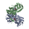
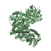
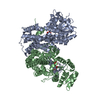
| Title | X-RAY STRUCTURE OF 6-PHOSPHOGLUCONATE DEHYDROGENASE FROM THE PROTOZOAN PARASITE T. BRUCEI | ||||||
 Components Components | 6-PHOSPHOGLUCONATE DEHYDROGENASE | ||||||
 Keywords Keywords | OXIDOREDUCTASE / CHOH(D)-NADP+(B) | ||||||
| Function / homology |  Function and homology information Function and homology informationD-gluconate metabolic process / phosphogluconate dehydrogenase (NADP+-dependent, decarboxylating) / phosphogluconate dehydrogenase (decarboxylating) activity / glycosome / pentose-phosphate shunt / ciliary plasm / NADP binding / cytoplasmSimilarity search - Function | ||||||
| Biological species |  Trypanosoma brucei (eukaryote) Trypanosoma brucei (eukaryote) | ||||||
| Method | X-RAY DIFFRACTION / MOLECULAR REPLACEMENT / Resolution: 2.82 Å | ||||||
 Authors Authors | Dohnalek, J. / Phillips, C. / Gover, S. / Barrett, M.P. / Adams, M.J. | ||||||
 Citation Citation | Journal: J.Mol.Biol. / Year: 1998 Title: A 2.8 A resolution structure of 6-phosphogluconate dehydrogenase from the protozoan parasite Trypanosoma brucei: comparison with the sheep enzyme accounts for differences in activity with ...Title: A 2.8 A resolution structure of 6-phosphogluconate dehydrogenase from the protozoan parasite Trypanosoma brucei: comparison with the sheep enzyme accounts for differences in activity with coenzyme and substrate analogues. Authors: Phillips, C. / Dohnalek, J. / Gover, S. / Barrett, M.P. / Adams, M.J. #1: Journal: Protein Expr.Purif. / Year: 1994Title: Overexpression in Escherichia Coli and Purification of the 6-Phosphogluconate Dehydrogenase of Trypanosoma Brucei Authors: Barrett, M.P. / Phillips, C. / Adams, M.J. / Le Page, R.W. #2: Journal: Structure / Year: 1994Title: Crystallographic Study of Coenzyme, Coenzyme Analogue and Substrate Binding in 6-Phosphogluconate Dehydrogenase: Implications for Nadp Specificity and the Enzyme Mechanism Authors: Adams, M.J. / Ellis, G.H. / Gover, S. / Naylor, C.E. / Phillips, C. #3: Journal: Structure / Year: 1994Title: Erratum. Crystallographic Study of Coenzyme, Coenzyme Analogue and Substrate Binding in 6-Phosphogluconate Dehydrogenase: Implications for Nadp Specificity and the Enzyme Mechanism Authors: Adams, M.J. / Ellis, G.H. / Gover, S. / Naylor, C.E. / Phillips, C. #4: Journal: J.Mol.Biol. / Year: 1993Title: Preliminary Crystallographic Study of 6-Phosphogluconate Dehydrogenase from Trypanosoma Brucei Authors: Phillips, C. / Barrett, M.P. / Gover, S. / Le Page, R.W. / Adams, M.J. | ||||||
| History |
|
- Structure visualization
| Structure viewer | Molecule: MolmilJmol/JSmol |
|---|
- Downloads & links
-Download
| PDBx/mmCIF format | 1pgj.cif.gz | 186.9 KB | Display | PDBx/mmCIF format |
|---|---|---|---|---|
| PDB format | pdb1pgj.ent.gz | 154.5 KB | Display | PDB format |
| PDBx/mmJSON format | 1pgj.json.gz | Tree view | PDBx/mmJSON format | |
| Others |  Other downloads Other downloads |
-Validation report
| Arichive directory | https://data.pdbj.org/pub/pdb/validation_reports/pg/1pgjftp://data.pdbj.org/pub/pdb/validation_reports/pg/1pgj | HTTPS FTP |
|---|
-Related structure data
| Related structure data |  2pgdS S: Starting model for refinement |
|---|---|
| Similar structure data |


-Links
 PDBj
PDBj


- Assembly
| Deposited unit | 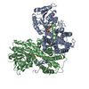
| ||||||||
|---|---|---|---|---|---|---|---|---|---|
| 1 |
| ||||||||
| Unit cell |
| ||||||||
| Components on special symmetry positions |
| ||||||||
| Noncrystallographic symmetry (NCS) | NCS oper: (Code: given Matrix: (0.48179, -0.85365, -0.19789), Vector : |
-Components
| #1: Protein | / 6PGDH / 6-PGDH Mass: 52091.539 Da / Num. of mol.: 2 Source method: isolated from a genetically manipulated source Source: (gene. exp.) Trypanosoma brucei (eukaryote) / Strain: BRUCEI / Cell line: BL21 / Gene: T. BRUCEI GND / Variant: TRUC427 / Plasmid: PT7GND / Species (production host): Escherichia coli / Gene (production host): T. BRUCEI GND / Production host:  Escherichia coli BL21(DE3) (bacteria) / Strain (production host): BL21 (DE3) Escherichia coli BL21(DE3) (bacteria) / Strain (production host): BL21 (DE3)References: UniProt: P31072, phosphogluconate dehydrogenase (NADP+-dependent, decarboxylating) #2: Chemical | ChemComp-SO4 / Sulfate  Mass: 96.063 Da / Num. of mol.: 4 / Source method: obtained synthetically / Formula: SO4 Mass: 96.063 Da / Num. of mol.: 4 / Source method: obtained synthetically / Formula: SO4#3: Water | ChemComp-HOH / | Water Mass: 18.015 Da / Num. of mol.: 129 / Source method: isolated from a natural source / Formula: H2O Mass: 18.015 Da / Num. of mol.: 129 / Source method: isolated from a natural source / Formula: H2O |
|---|
-Experimental details
-Experiment
| Experiment | Method: X-RAY DIFFRACTION / Number of used crystals: 1 |
|---|
- Sample preparation
| Crystal | Density Matthews: 2.96 Å3/Da / Density % sol: 58.5 % Description: DATA COLLECTED BY OSCILLATION METHOD IN STEPS OF 1 DEGREE IN PHI. R SYM GIVEN IS FOR I > 4 SIG(I). |
|---|---|
| Crystal grow | Method: vapor diffusion, hanging drop / pH: 7 Details: CRYSTALLISED FROM HANGING DROP WHICH ALSO CONTAINED 50MM POTASSIUM PHOSPHATE, 5MM DTT AND 30% SATURATED AMMONIUM SULPHATE, PH 7.0. THE WELL SOLUTION WAS 45% SATURATED AMMONIUM SULPHATE., ...Details: CRYSTALLISED FROM HANGING DROP WHICH ALSO CONTAINED 50MM POTASSIUM PHOSPHATE, 5MM DTT AND 30% SATURATED AMMONIUM SULPHATE, PH 7.0. THE WELL SOLUTION WAS 45% SATURATED AMMONIUM SULPHATE., vapor diffusion - hanging drop |
| Crystal grow | *PLUS Details: Barrett, M.P., (1994) Protein Expr. Purif., 5, 44. |
-Data collection
| Diffraction | Mean temperature: 293 K |
|---|---|
| Diffraction source | Source: ROTATING ANODE / Type: RIGAKU RUH2R / Wavelength: 1.5418 |
| Detector | Type: MARRESEARCH / Detector: IMAGE PLATE / Date: Nov 1, 1992 / Details: COLLIMATOR, DUAL SLITS |
| Radiation | Monochromator: GRAPHITE(002) / Monochromatic (M) / Laue (L): M / Scattering type: x-ray |
| Radiation wavelength | Wavelength: 1.5418 Å / Relative weight: 1 |
| Reflection | Resolution: 2.8→20 Å / Num. obs: 29373 / % possible obs: 95.9 % / Redundancy: 7.2 % / Rsym value: 0.099 / Net I/σ(I): 17.9 |
| Reflection | *PLUS Observed criterion σ(I): 4 / Num. measured all: 212041 / Rmerge(I) obs: 0.099 |
- Processing
| Software |
| ||||||||||||||||||||||||||||||||||||||||||||||||||||||||||||||||||||||||||||||||
|---|---|---|---|---|---|---|---|---|---|---|---|---|---|---|---|---|---|---|---|---|---|---|---|---|---|---|---|---|---|---|---|---|---|---|---|---|---|---|---|---|---|---|---|---|---|---|---|---|---|---|---|---|---|---|---|---|---|---|---|---|---|---|---|---|---|---|---|---|---|---|---|---|---|---|---|---|---|---|---|---|---|
| Refinement | Method to determine structure: MOLECULAR REPLACEMENT Starting model: SHEEP DIMER - SEE PDB ENTRY 2PGD Resolution: 2.82→19.76 Å / Data cutoff low absF: 0 Isotropic thermal model: TARGET SIGMA FOR 1-2 B FA PAIRS (BOND), 1-3 PAIRS (ANGLE) Cross valid method: SEE JRNL REFERENCE / σ(F): 0 Details: FOR DETAILS OF RESTRAINED RESIDUES AND PARAMETERS USED IN BULK SOLVENT MODELLING, SEE JRNL REFERENCE. COORDINATES GIVEN ARE THOSE AFTER A FINAL NON-PARTITIONED (I.E., ALL DATA) REFINEMENT ...Details: FOR DETAILS OF RESTRAINED RESIDUES AND PARAMETERS USED IN BULK SOLVENT MODELLING, SEE JRNL REFERENCE. COORDINATES GIVEN ARE THOSE AFTER A FINAL NON-PARTITIONED (I.E., ALL DATA) REFINEMENT CYCLE, FOR WHICH R = 0.188 AND BIN R = 0.267.
| ||||||||||||||||||||||||||||||||||||||||||||||||||||||||||||||||||||||||||||||||
| Displacement parameters | Biso mean: 26.5 Å2 | ||||||||||||||||||||||||||||||||||||||||||||||||||||||||||||||||||||||||||||||||
| Refine analyze | Luzzati coordinate error obs: 0.3 Å | ||||||||||||||||||||||||||||||||||||||||||||||||||||||||||||||||||||||||||||||||
| Refinement step | Cycle: LAST / Resolution: 2.82→19.76 Å
| ||||||||||||||||||||||||||||||||||||||||||||||||||||||||||||||||||||||||||||||||
| Refine LS restraints |
| ||||||||||||||||||||||||||||||||||||||||||||||||||||||||||||||||||||||||||||||||
| Refine LS restraints NCS | NCS model details: RESTRAINTS / Rms dev Biso : 2.14 Å2 / Rms dev position: 0.1 Å / Weight Biso : 2 / Weight position: 80 | ||||||||||||||||||||||||||||||||||||||||||||||||||||||||||||||||||||||||||||||||
| LS refinement shell | Resolution: 2.82→2.93 Å / Total num. of bins used: 8
| ||||||||||||||||||||||||||||||||||||||||||||||||||||||||||||||||||||||||||||||||
| Xplor file |
|
