 Movie
Movie Controller

[English] 日本語
 Yorodumi
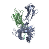
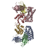
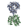
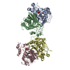
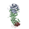
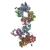
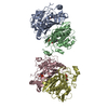
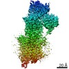
- PDB-1p4l: Crystal structure of NK receptor Ly49C mutant with its MHC class ... -
+ Open data

- Basic information
| Entry | Database: PDB / ID: 1p4l | ||||||
|---|---|---|---|---|---|---|---|
| Title | Crystal structure of NK receptor Ly49C mutant with its MHC class I ligand H-2Kb | ||||||
 Components Components |
| ||||||
 Keywords Keywords |  IMMUNE SYSTEM / Natural Killer Receptor / MHC class I / C-type Lectin-like domain IMMUNE SYSTEM / Natural Killer Receptor / MHC class I / C-type Lectin-like domain | ||||||
| Function / homology |  Function and homology information Function and homology informationintracellular amino acid homeostasis / RUNX1 regulates transcription of genes involved in differentiation of keratinocytes / phagolysosome / monoatomic ion homeostasis / TAP1 binding / TAP2 binding / positive regulation of antibody-dependent cellular cytotoxicity / regulation of natural killer cell mediated immunity / positive regulation of TRAIL production / negative regulation of natural killer cell activation ...intracellular amino acid homeostasis / RUNX1 regulates transcription of genes involved in differentiation of keratinocytes / phagolysosome / monoatomic ion homeostasis / TAP1 binding / TAP2 binding / positive regulation of antibody-dependent cellular cytotoxicity / regulation of natural killer cell mediated immunity / positive regulation of TRAIL production / negative regulation of natural killer cell activation / antigen processing and presentation of exogenous peptide antigen via MHC class Ib / MHC class Ib protein complex / positive regulation of natural killer cell mediated immunity / positive regulation of natural killer cell cytokine production / positive regulation of natural killer cell activation / natural killer cell tolerance induction / natural killer cell lectin-like receptor binding / cis-Golgi network membrane / response to steroid hormone / embryo development ending in birth or egg hatching / Endosomal/Vacuolar pathway / DAP12 interactions / Antigen Presentation: Folding, assembly and peptide loading of class I MHC / ER-Phagosome pathway / DAP12 signaling / negative regulation of natural killer cell mediated cytotoxicity / Immunoregulatory interactions between a Lymphoid and a non-Lymphoid cell / positive regulation of interleukin-13 production / response to corticosterone / positive regulation of natural killer cell mediated cytotoxicity / regulation of membrane depolarization / inner ear development / positive regulation of natural killer cell proliferation / T cell mediated cytotoxicity directed against tumor cell target / antigen processing and presentation of endogenous peptide antigen via MHC class I via ER pathway, TAP-dependent / positive regulation of memory T cell activation / positive regulation of immunoglobulin production / TAP complex binding / antigen processing and presentation of exogenous peptide antigen via MHC class I / Golgi medial cisterna / positive regulation of CD8-positive, alpha-beta T cell activation / CD8-positive, alpha-beta T cell activation / positive regulation of interleukin-4 production / positive regulation of CD8-positive, alpha-beta T cell proliferation / CD8 receptor binding / MHC class I protein binding / cellular defense response / endoplasmic reticulum exit site / beta-2-microglobulin binding / phagocytic vesicle / monoatomic ion transport / TAP binding / negative regulation of T cell proliferation / protection from natural killer cell mediated cytotoxicity / antigen processing and presentation of endogenous peptide antigen via MHC class I via ER pathway, TAP-independent / antigen processing and presentation of endogenous peptide antigen via MHC class Ib / detection of bacterium / Neutrophil degranulation / T cell receptor binding / 14-3-3 protein binding / response to progesterone / early endosome lumen / Neutrophil degranulation / lumenal side of endoplasmic reticulum membrane / peptide binding / antigen processing and presentation of exogenous protein antigen via MHC class Ib, TAP-dependent / cellular response to iron(III) ion / negative regulation of forebrain neuron differentiation / response to molecule of bacterial origin / regulation of erythrocyte differentiation / regulation of iron ion transport / MHC class I peptide loading complex / HFE-transferrin receptor complex / T cell mediated cytotoxicity / cellular response to iron ion / antigen processing and presentation of endogenous peptide antigen via MHC class I / positive regulation of T cell cytokine production / MHC class I protein complex / multicellular organismal-level iron ion homeostasis / positive regulation of T cell mediated cytotoxicity / peptide antigen assembly with MHC class II protein complex / negative regulation of neurogenesis / positive regulation of receptor-mediated endocytosis / MHC class II protein complex / cellular response to nicotine / response to estrogen / phagocytic vesicle membrane / peptide antigen binding / positive regulation of cellular senescence / antigen processing and presentation of exogenous peptide antigen via MHC class II / positive regulation of immune response / negative regulation of epithelial cell proliferation / positive regulation of T cell activation / positive regulation of type II interferon production / antimicrobial humoral immune response mediated by antimicrobial peptide / sensory perception of smell / negative regulation of neuron projection development / positive regulation of tumor necrosis factor production / MHC class II protein complex binding / late endosome membraneSimilarity search - Function | ||||||
| Biological species |  Mus musculus (house mouse) Mus musculus (house mouse) | ||||||
| Method | X-RAY DIFFRACTION / SYNCHROTRON / MOLECULAR REPLACEMENT / Resolution: 2.9 Å | ||||||
 Authors Authors | Dam, J. / Guan, R. / Natarajan, K. / Dimasi, N. / Mariuzza, R.A. | ||||||
 Citation Citation | Journal: Nat.Immunol. / Year: 2003 Title: Variable MHC class I engagement by Ly49 natural killer cell receptors demonstrated by the crystal structure of Ly49C bound to H-2K(b). Authors: Dam, J. / Guan, R. / Natarajan, K. / Dimasi, N. / Chlewicki, L.K. / Kranz, D.M. / Schuck, P. / Margulies, D.H. / Mariuzza, R.A. | ||||||
| History |
|
- Structure visualization
| Structure viewer | Molecule: MolmilJmol/JSmol |
|---|
- Downloads & links
-Download
| PDBx/mmCIF format | 1p4l.cif.gz | 107.8 KB | Display | PDBx/mmCIF format |
|---|---|---|---|---|
| PDB format | pdb1p4l.ent.gz | 83.9 KB | Display | PDB format |
| PDBx/mmJSON format | 1p4l.json.gz | Tree view | PDBx/mmJSON format | |
| Others |  Other downloads Other downloads |
-Validation report
| Arichive directory | https://data.pdbj.org/pub/pdb/validation_reports/p4/1p4lftp://data.pdbj.org/pub/pdb/validation_reports/p4/1p4l | HTTPS FTP |
|---|
-Related structure data
| Related structure data | 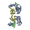 1p1zC  1qo3S 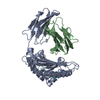 2vabS C: citing same article ( S: Starting model for refinement |
|---|---|
| Similar structure data |


-Links
 PDBj
PDBj


- Assembly
| Deposited unit | 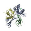
| ||||||||
|---|---|---|---|---|---|---|---|---|---|
| 1 | 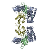
| ||||||||
| Unit cell |
|
-Components
| #1: Protein | Mass: 31648.322 Da / Num. of mol.: 1 / Fragment: extracellular alpha-1, alpha-2, alpha-3 Source method: isolated from a genetically manipulated source Source: (gene. exp.) Mus musculus (house mouse) / Gene: H2-K / Production host:  Escherichia coli (E. coli) / References: UniProt: P01901 Escherichia coli (E. coli) / References: UniProt: P01901 |
|---|---|
| #2: Protein | Beta-2 microglobulin Mass: 11660.350 Da / Num. of mol.: 1 Source method: isolated from a genetically manipulated source Source: (gene. exp.) Mus musculus (house mouse) / Gene: B2M / Production host: Escherichia coli (E. coli) / References: UniProt: P01887 |
| #3: Protein/peptide | Mass: 964.137 Da / Num. of mol.: 1 / Source method: obtained synthetically Details: The peptide was chemically synthesized. The sequence of the peptide is naturally found in Gallus gallus (chicken). References: UniProt: P01012 |
| #4: Protein | Mass: 14556.108 Da / Num. of mol.: 1 / Fragment: C-Type Lectin-Like Domain / Mutation: S171G, E193G, R223K Source method: isolated from a genetically manipulated source Source: (gene. exp.) Mus musculus (house mouse) / Gene: KLRA3 OR LY49C OR LY-49C / Production host: Escherichia coli (E. coli) / References: UniProt: Q64329 |
-Experimental details
-Experiment
| Experiment | Method: X-RAY DIFFRACTION / Number of used crystals: 1 |
|---|
- Sample preparation
| Crystal | Density Matthews: 3.17 Å3/Da / Density % sol: 61.24 % | ||||||||||||||||||||||||||||||
|---|---|---|---|---|---|---|---|---|---|---|---|---|---|---|---|---|---|---|---|---|---|---|---|---|---|---|---|---|---|---|---|
| Crystal grow | Temperature: 298 K / Method: vapor diffusion, hanging drop / pH: 7.5 Details: Protein 8mg/ml, 0.1 M HEPES-Na pH 7.5, 2% v/v PEG400, 2.0 M Ammonium Sulfate, VAPOR DIFFUSION, HANGING DROP, temperature 298K | ||||||||||||||||||||||||||||||
| Crystal grow | *PLUS Method: vapor diffusion, hanging drop | ||||||||||||||||||||||||||||||
| Components of the solutions | *PLUS
|
-Data collection
| Diffraction | Mean temperature: 100 K |
|---|---|
| Diffraction source | Source: SYNCHROTRON / Site: CHESS  / Beamline: F1 / Wavelength: 0.916 Å / Beamline: F1 / Wavelength: 0.916 Å |
| Detector | Type: ADSC QUANTUM 4 / Detector: CCD / Date: Oct 31, 2002 |
| Radiation | Monochromator: Si 111 CHANNEL / Protocol: SINGLE WAVELENGTH / Monochromatic (M) / Laue (L): M / Scattering type: x-ray |
| Radiation wavelength | Wavelength: 0.916 Å / Relative weight: 1 |
| Reflection | Resolution: 2.9→19.6 Å / Num. all: 16931 / Num. obs: 16931 / % possible obs: 97.5 % / Observed criterion σ(F): 0 / Observed criterion σ(I): 0 / Biso Wilson estimate: 82.2 Å2 |
| Reflection shell | Resolution: 2.9→2.95 Å / Rmerge(I) obs: 0.325 / Num. unique all: 840 / % possible all: 97.9 |
| Reflection | *PLUS Highest resolution: 2.9 Å / Rmerge(I) obs: 0.062 |
| Reflection shell | *PLUS % possible obs: 97.9 % / Num. unique obs: 840 |
- Processing
| Software |
| ||||||||||||||||||||||||||||||||||||||||||||||||||||||||||||
|---|---|---|---|---|---|---|---|---|---|---|---|---|---|---|---|---|---|---|---|---|---|---|---|---|---|---|---|---|---|---|---|---|---|---|---|---|---|---|---|---|---|---|---|---|---|---|---|---|---|---|---|---|---|---|---|---|---|---|---|---|---|
| Refinement | Method to determine structure: MOLECULAR REPLACEMENT Starting model: PDB ENTRY 2VAB and 1QO3 Resolution: 2.9→19.96 Å / Isotropic thermal model: RESTRAINED / Cross valid method: THROUGHOUT / σ(F): 0 / Stereochemistry target values: Engh & Huber
| ||||||||||||||||||||||||||||||||||||||||||||||||||||||||||||
| Solvent computation | Solvent model: FLAT MODEL / Bsol: 53.1893 Å2 / ksol: 0.356108 e/Å3 | ||||||||||||||||||||||||||||||||||||||||||||||||||||||||||||
| Displacement parameters | Biso mean: 76 Å2
| ||||||||||||||||||||||||||||||||||||||||||||||||||||||||||||
| Refine analyze | Luzzati coordinate error free: 0.53 Å / Luzzati sigma a free: 0.67 Å | ||||||||||||||||||||||||||||||||||||||||||||||||||||||||||||
| Refinement step | Cycle: LAST / Resolution: 2.9→19.96 Å
| ||||||||||||||||||||||||||||||||||||||||||||||||||||||||||||
| Refine LS restraints |
| ||||||||||||||||||||||||||||||||||||||||||||||||||||||||||||
| LS refinement shell | Resolution: 2.9→3.08 Å / Total num. of bins used: 6
| ||||||||||||||||||||||||||||||||||||||||||||||||||||||||||||
| Xplor file |
| ||||||||||||||||||||||||||||||||||||||||||||||||||||||||||||
| Refinement | *PLUS Lowest resolution: 20 Å / % reflection Rfree: 4.5 % | ||||||||||||||||||||||||||||||||||||||||||||||||||||||||||||
| Solvent computation | *PLUS | ||||||||||||||||||||||||||||||||||||||||||||||||||||||||||||
| Displacement parameters | *PLUS | ||||||||||||||||||||||||||||||||||||||||||||||||||||||||||||
| Refine LS restraints | *PLUS
|
