 Movie
Movie Controller

[English] 日本語
 Yorodumi
- PDB-1p39: DC26 MUTANT OF VACCINIA VIRUS PROTEIN VP39 IN COMPLEX WITH S-ADEN... -
+ Open data

- Basic information
| Entry | Database: PDB / ID: 1p39 | |||||||||
|---|---|---|---|---|---|---|---|---|---|---|
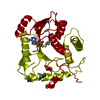
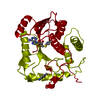
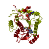
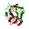
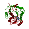
| Title | DC26 MUTANT OF VACCINIA VIRUS PROTEIN VP39 IN COMPLEX WITH S-ADENOSYLHOMOCYSTEINE AND M7G(5')PPPG | |||||||||
 Components Components | VP39 | |||||||||
 Keywords Keywords |  METHYLTRANSFERASE / RNA CAP / POLY(A) POLYMERASE / VACCINIA / MRNA PROCESSING / TRANSCRIPTION METHYLTRANSFERASE / RNA CAP / POLY(A) POLYMERASE / VACCINIA / MRNA PROCESSING / TRANSCRIPTION | |||||||||
| Function / homology |  Function and homology information Function and homology informationregulation of mRNA 3'-end processing / 7-methylguanosine mRNA capping / translation elongation factor activity / virion component / methyltransferase cap1 / mRNA (nucleoside-2'-O-)-methyltransferase activity / RNA bindingSimilarity search - Function | |||||||||
| Biological species |  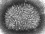 Vaccinia virus Vaccinia virus | |||||||||
| Method | X-RAY DIFFRACTION / MOLECULAR REPLACEMENT / Resolution: 2 Å | |||||||||
 Authors Authors | Hodel, A.E. / Gershon, P.D. / Quiocho, F.A. | |||||||||
 Citation Citation | Journal: Nat.Struct.Biol. / Year: 1997 Title: Specific protein recognition of an mRNA cap through its alkylated base. Authors: Hodel, A.E. / Gershon, P.D. / Shi, X. / Wang, S.M. / Quiocho, F.A. #1: Journal: Cell(Cambridge,Mass.) / Year: 1996Title: The 1.85 A Structure of Vaccinia Protein Vp39: A Bifunctional Enzyme that Participates in the Modification of Both Mrna Ends Authors: Hodel, A.E. / Gershon, P.D. / Shi, X. / Quiocho, F.A. | |||||||||
| History |
|
- Structure visualization
| Structure viewer | Molecule: MolmilJmol/JSmol |
|---|
- Downloads & links
-Download
| PDBx/mmCIF format | 1p39.cif.gz | 75.6 KB | Display | PDBx/mmCIF format |
|---|---|---|---|---|
| PDB format | pdb1p39.ent.gz | 58.3 KB | Display | PDB format |
| PDBx/mmJSON format | 1p39.json.gz | Tree view | PDBx/mmJSON format | |
| Others |  Other downloads Other downloads |
-Validation report
| Arichive directory | https://data.pdbj.org/pub/pdb/validation_reports/p3/1p39ftp://data.pdbj.org/pub/pdb/validation_reports/p3/1p39 | HTTPS FTP |
|---|
-Related structure data
| Related structure data |  1v39C  1vp3C  1vp9C  2vp3C 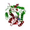 1vptS S: Starting model for refinement C: citing same article ( |
|---|---|
| Similar structure data |


-Links
 PDBj
PDBj
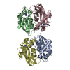
- Assembly
| Deposited unit | 
| ||||||||
|---|---|---|---|---|---|---|---|---|---|
| 1 | 
| ||||||||
| Unit cell |
| ||||||||
| Components on special symmetry positions |
|
-Components
| #1: Protein | Mass: 37133.703 Da / Num. of mol.: 1 / Mutation: 26 C-TERMINAL RESIDUES DELETED Source method: isolated from a genetically manipulated source Source: (gene. exp.) Vaccinia virus / Genus: Orthopoxvirus / Strain: WR / Cell line: BL21 / Plasmid: BL21 / Species (production host): Escherichia coli / Production host:  Escherichia coli BL21(DE3) (bacteria) / Strain (production host): BL21 (DE3) Escherichia coli BL21(DE3) (bacteria) / Strain (production host): BL21 (DE3)References: UniProt: P07617, polynucleotide adenylyltransferase |
|---|---|
| #2: Chemical | ChemComp-SAH / S-Adenosyl-L-homocysteine  Type: L-peptide linking / Mass: 384.411 Da / Num. of mol.: 1 / Source method: obtained synthetically / Formula: C14H20N6O5S Type: L-peptide linking / Mass: 384.411 Da / Num. of mol.: 1 / Source method: obtained synthetically / Formula: C14H20N6O5S |
| #3: Chemical | ChemComp-MGT /   Mass: 539.223 Da / Num. of mol.: 1 / Source method: obtained synthetically / Formula: C11H20N5O14P3 Mass: 539.223 Da / Num. of mol.: 1 / Source method: obtained synthetically / Formula: C11H20N5O14P3 |
| #4: Water | ChemComp-HOH / Water Mass: 18.015 Da / Num. of mol.: 205 / Source method: isolated from a natural source / Formula: H2O Mass: 18.015 Da / Num. of mol.: 205 / Source method: isolated from a natural source / Formula: H2O |
-Experimental details
-Experiment
| Experiment | Method: X-RAY DIFFRACTION / Number of used crystals: 1 |
|---|
- Sample preparation
| Crystal | Density Matthews: 2.71 Å3/Da / Density % sol: 53 % | ||||||||||||||||||||||||
|---|---|---|---|---|---|---|---|---|---|---|---|---|---|---|---|---|---|---|---|---|---|---|---|---|---|
| Crystal grow | pH: 4.5 Details: DC26 CRYSTALLIZED IN 2MM ADOHCY AND 2MM M7GPPPG., pH 4.5 | ||||||||||||||||||||||||
| Crystal grow | *PLUS Details: macro-seeding, Hodel, A.E., (1996) Cell(Cambridge,Mass.), 85, 247.Method: other | ||||||||||||||||||||||||
| Components of the solutions | *PLUS
|
-Data collection
| Diffraction | Mean temperature: 103 K |
|---|---|
| Diffraction source | Source: ROTATING ANODE / Type: RIGAKU RUH2R / Wavelength: 1.5418 |
| Detector | Type: MACSCIENCE / Detector: IMAGE PLATE / Date: Apr 27, 1996 / Details: MIRROR |
| Radiation | Monochromator: NI FILTER / Monochromatic (M) / Laue (L): M / Scattering type: x-ray |
| Radiation wavelength | Wavelength: 1.5418 Å / Relative weight: 1 |
| Reflection | Resolution: 1.9→15 Å / Num. obs: 30508 / % possible obs: 96 % / Observed criterion σ(I): 2 / Redundancy: 4 % / Rmerge(I) obs: 0.049 / Net I/σ(I): 35 |
| Reflection shell | Resolution: 1.9→1.97 Å / Redundancy: 3 % / Rmerge(I) obs: 0.31 / Mean I/σ(I) obs: 5 / % possible all: 89 |
| Reflection shell | *PLUS % possible obs: 89 % |
- Processing
| Software |
| ||||||||||||||||||||||||||||||||||||||||||||||||||||||||||||||||||||||||||||||||
|---|---|---|---|---|---|---|---|---|---|---|---|---|---|---|---|---|---|---|---|---|---|---|---|---|---|---|---|---|---|---|---|---|---|---|---|---|---|---|---|---|---|---|---|---|---|---|---|---|---|---|---|---|---|---|---|---|---|---|---|---|---|---|---|---|---|---|---|---|---|---|---|---|---|---|---|---|---|---|---|---|---|
| Refinement | Method to determine structure: MOLECULAR REPLACEMENT Starting model: PDB ENTRY 1VPT Resolution: 2→8 Å / Data cutoff high absF: 100000 / Data cutoff low absF: 0.0001 / Isotropic thermal model: RESTRAINED / σ(F): 2
| ||||||||||||||||||||||||||||||||||||||||||||||||||||||||||||||||||||||||||||||||
| Displacement parameters | Biso mean: 31 Å2 | ||||||||||||||||||||||||||||||||||||||||||||||||||||||||||||||||||||||||||||||||
| Refinement step | Cycle: LAST / Resolution: 2→8 Å
| ||||||||||||||||||||||||||||||||||||||||||||||||||||||||||||||||||||||||||||||||
| Refine LS restraints |
| ||||||||||||||||||||||||||||||||||||||||||||||||||||||||||||||||||||||||||||||||
| Software | *PLUS Name: X-PLOR / Version: 3.1 / Classification: refinement | ||||||||||||||||||||||||||||||||||||||||||||||||||||||||||||||||||||||||||||||||
| Refinement | *PLUS | ||||||||||||||||||||||||||||||||||||||||||||||||||||||||||||||||||||||||||||||||
| Solvent computation | *PLUS | ||||||||||||||||||||||||||||||||||||||||||||||||||||||||||||||||||||||||||||||||
| Displacement parameters | *PLUS |
