 Movie
Movie Controller

[English] 日本語
 Yorodumi
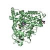
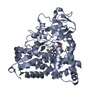
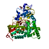
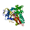
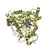
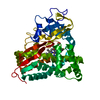
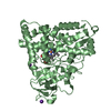
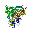
- PDB-1p2y: CRYSTAL STRUCTURE OF CYTOCHROME P450CAM IN COMPLEX WITH (S)-(-)-N... -
+ Open data

- Basic information
| Entry | Database: PDB / ID: 1p2y | ||||||
|---|---|---|---|---|---|---|---|
| Title | CRYSTAL STRUCTURE OF CYTOCHROME P450CAM IN COMPLEX WITH (S)-(-)-NICOTINE | ||||||
 Components Components | Cytochrome P450-cam | ||||||
 Keywords Keywords |  OXIDOREDUCTASE / MONOOXYGENASE / HEME / NICOTINE OXIDOREDUCTASE / MONOOXYGENASE / HEME / NICOTINE | ||||||
| Function / homology |  Function and homology informationcamphor 5-monooxygenase / camphor 5-monooxygenase activity / (+)-camphor catabolic process / iron ion binding / heme binding / cytoplasm Function and homology informationcamphor 5-monooxygenase / camphor 5-monooxygenase activity / (+)-camphor catabolic process / iron ion binding / heme binding / cytoplasmSimilarity search - Function | ||||||
| Biological species |  Pseudomonas putida (bacteria) Pseudomonas putida (bacteria) | ||||||
| Method | X-RAY DIFFRACTION / SYNCHROTRON / MOLECULAR REPLACEMENT / Resolution: 2.3 Å | ||||||
 Authors Authors | Strickler, M. / Goldstein, B.M. / Maxfield, K. / Shireman, L. / Kim, G. / Matteson, D. / Jones, J.P. | ||||||
 Citation Citation | Journal: Biochemistry / Year: 2003 Title: Crystallographic Studies on the Complex Behavior of Nicotine Binding to P450cam (CYP101)(dagger). Authors: Strickler, M. / Goldstein, B.M. / Maxfield, K. / Shireman, L. / Kim, G. / Matteson, D. / Jones, J.P. | ||||||
| History |
|
- Structure visualization
| Structure viewer | Molecule: MolmilJmol/JSmol |
|---|
- Downloads & links
-Download
| PDBx/mmCIF format | 1p2y.cif.gz | 90.7 KB | Display | PDBx/mmCIF format |
|---|---|---|---|---|
| PDB format | pdb1p2y.ent.gz | 68.9 KB | Display | PDB format |
| PDBx/mmJSON format | 1p2y.json.gz | Tree view | PDBx/mmJSON format | |
| Others |  Other downloads Other downloads |
-Validation report
| Arichive directory | https://data.pdbj.org/pub/pdb/validation_reports/p2/1p2yftp://data.pdbj.org/pub/pdb/validation_reports/p2/1p2y | HTTPS FTP |
|---|
-Related structure data


-Links
 PDBj
PDBj
- Assembly
| Deposited unit | 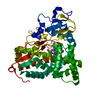
| ||||||||
|---|---|---|---|---|---|---|---|---|---|
| 1 |
| ||||||||
| Unit cell |
|
-Components
| #1: Protein | Mass: 47417.750 Da / Num. of mol.: 1 Source method: isolated from a genetically manipulated source Source: (gene. exp.) Pseudomonas putida (bacteria) / Gene: CAMC OR CYP101 / Plasmid: pBLUESCRIPT / Production host: Escherichia coli (E. coli) / Strain (production host): DH5ALPHA / References: UniProt: P00183, camphor 5-monooxygenase |
|---|---|
| #2: Chemical | ChemComp-HEM / Heme B  Mass: 616.487 Da / Num. of mol.: 1 / Source method: obtained synthetically / Formula: C34H32FeN4O4 Mass: 616.487 Da / Num. of mol.: 1 / Source method: obtained synthetically / Formula: C34H32FeN4O4 |
| #3: Chemical | ChemComp-NCT / (Nicotine  Mass: 162.232 Da / Num. of mol.: 1 / Source method: obtained synthetically / Formula: C10H14N2 / Comment: alkaloid*YM Mass: 162.232 Da / Num. of mol.: 1 / Source method: obtained synthetically / Formula: C10H14N2 / Comment: alkaloid*YM |
| #4: Water | ChemComp-HOH / Water Mass: 18.015 Da / Num. of mol.: 122 / Source method: isolated from a natural source / Formula: H2O Mass: 18.015 Da / Num. of mol.: 122 / Source method: isolated from a natural source / Formula: H2O |
-Experimental details
-Experiment
| Experiment | Method: X-RAY DIFFRACTION / Number of used crystals: 1 |
|---|
- Sample preparation
| Crystal | Density Matthews: 2.7 Å3/Da / Density % sol: 54.11 % | |||||||||||||||
|---|---|---|---|---|---|---|---|---|---|---|---|---|---|---|---|---|
| Crystal grow | Temperature: 277 K / Method: vapor diffusion, sitting drop / pH: 7 Details: PEG 8000, potassium phosphate, potassium chloride, dithiothreitol, nicotine, pH 7.0, VAPOR DIFFUSION, SITTING DROP, temperature 277K | |||||||||||||||
| Crystal grow | *PLUS Temperature: 4 ℃ / Method: vapor diffusion, sitting drop | |||||||||||||||
| Components of the solutions | *PLUS
|
-Data collection
| Diffraction | Mean temperature: 103 K |
|---|---|
| Diffraction source | Source: SYNCHROTRON / Site: CHESS  / Beamline: A1 / Wavelength: 1 Å / Beamline: A1 / Wavelength: 1 Å |
| Detector | Type: ADSC QUANTUM 1 / Detector: CCD / Date: Jul 7, 1996 |
| Radiation | Monochromator: Si (III) / Protocol: SINGLE WAVELENGTH / Monochromatic (M) / Laue (L): M / Scattering type: x-ray |
| Radiation wavelength | Wavelength: 1 Å / Relative weight: 1 |
| Reflection | Resolution: 2.3→39.52 Å / Num. all: 25149 / Num. obs: 21616 / % possible obs: 86 % / Observed criterion σ(I): -3 / Redundancy: 4.7 % / Biso Wilson estimate: 13 Å2 / Rsym value: 0.066 / Net I/σ(I): 12.4 |
| Reflection shell | Resolution: 2.3→2.32 Å / Mean I/σ(I) obs: 4 / Num. unique all: 334 / Rsym value: 0.242 / % possible all: 56.4 |
| Reflection | *PLUS Rmerge(I) obs: 0.066 |
| Reflection shell | *PLUS % possible obs: 56.4 % / Rmerge(I) obs: 0.242 |
- Processing
| Software |
| ||||||||||||||||||||||||||||||||||||
|---|---|---|---|---|---|---|---|---|---|---|---|---|---|---|---|---|---|---|---|---|---|---|---|---|---|---|---|---|---|---|---|---|---|---|---|---|---|
| Refinement | Method to determine structure: MOLECULAR REPLACEMENT / Resolution: 2.3→39.52 Å / Rfactor Rfree error: 0.006 / Data cutoff high absF: 441226.16 / Data cutoff high rms absF: 441226.16 / Data cutoff low absF: 0 / Isotropic thermal model: RESTRAINED / Cross valid method: THROUGHOUT / σ(F): 0 / Stereochemistry target values: Engh & Huber / Details: BULK SOLVENT MODEL USED
| ||||||||||||||||||||||||||||||||||||
| Solvent computation | Solvent model: FLAT MODEL / Bsol: 13.9252 Å2 / ksol: 0.321998 e/Å3 | ||||||||||||||||||||||||||||||||||||
| Displacement parameters | Biso mean: 23.2 Å2
| ||||||||||||||||||||||||||||||||||||
| Refine analyze |
| ||||||||||||||||||||||||||||||||||||
| Refinement step | Cycle: LAST / Resolution: 2.3→39.52 Å
| ||||||||||||||||||||||||||||||||||||
| Refine LS restraints |
| ||||||||||||||||||||||||||||||||||||
| LS refinement shell | Resolution: 2.3→2.44 Å / Rfactor Rfree error: 0.018 / Total num. of bins used: 6
| ||||||||||||||||||||||||||||||||||||
| Xplor file |
| ||||||||||||||||||||||||||||||||||||
| Refinement | *PLUS Highest resolution: 2.3 Å / Rfactor Rfree: 0.264 | ||||||||||||||||||||||||||||||||||||
| Solvent computation | *PLUS | ||||||||||||||||||||||||||||||||||||
| Displacement parameters | *PLUS | ||||||||||||||||||||||||||||||||||||
| Refine LS restraints | *PLUS
|
