 Movie
Movie Controller


+ Open data

- Basic information
| Entry | Database: PDB / ID: 1ow3 | ||||||
|---|---|---|---|---|---|---|---|
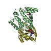
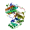
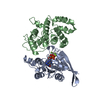
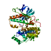
| Title | Crystal Structure of RhoA.GDP.MgF3-in Complex with RhoGAP | ||||||
 Components Components |
| ||||||
 Keywords Keywords | GENE REGULATION/SIGNALING PROTEIN /  Complex / GTPase / GAP / transition state / GENE REGULATION-SIGNALING PROTEIN COMPLEX Complex / GTPase / GAP / transition state / GENE REGULATION-SIGNALING PROTEIN COMPLEX | ||||||
| Function / homology |  Function and homology information Function and homology informationnegative regulation of endocytic recycling / transferrin transport / aortic valve formation / alpha-beta T cell lineage commitment / mitotic cleavage furrow formation / bone trabecula morphogenesis / positive regulation of lipase activity / endothelial tube lumen extension / skeletal muscle satellite cell migration / positive regulation of vascular associated smooth muscle contraction ...negative regulation of endocytic recycling / transferrin transport / aortic valve formation / alpha-beta T cell lineage commitment / mitotic cleavage furrow formation / bone trabecula morphogenesis / positive regulation of lipase activity / endothelial tube lumen extension / skeletal muscle satellite cell migration / positive regulation of vascular associated smooth muscle contraction / angiotensin-mediated vasoconstriction involved in regulation of systemic arterial blood pressure / SLIT2:ROBO1 increases RHOA activity / RHO GTPases Activate Rhotekin and Rhophilins / Roundabout signaling pathway / negative regulation of intracellular steroid hormone receptor signaling pathway / Axonal growth inhibition (RHOA activation) / Axonal growth stimulation / regulation of neural precursor cell proliferation / cleavage furrow formation / regulation of modification of postsynaptic actin cytoskeleton / regulation of osteoblast proliferation / forebrain radial glial cell differentiation / cell junction assembly / apical junction assembly / regulation of systemic arterial blood pressure by endothelin / cellular response to chemokine / negative regulation of cell migration involved in sprouting angiogenesis / beta selection / establishment of epithelial cell apical/basal polarity / regulation of modification of postsynaptic structure / negative regulation of cell size / RHO GTPases Activate ROCKs / negative regulation of oxidative phosphorylation / negative regulation of motor neuron apoptotic process / RHOF GTPase cycle / RHO GTPases activate CIT / RHOD GTPase cycle / PCP/CE pathway / Sema4D induced cell migration and growth-cone collapse / RHO GTPases activate KTN1 / apolipoprotein A-I-mediated signaling pathway / positive regulation of podosome assembly / negative regulation of cell-substrate adhesion / Wnt signaling pathway, planar cell polarity pathway / regulation of small GTPase mediated signal transduction / Sema4D mediated inhibition of cell attachment and migration / ossification involved in bone maturation / positive regulation of alpha-beta T cell differentiation / odontogenesis / motor neuron apoptotic process / wound healing, spreading of cells / RND2 GTPase cycle / PI3K/AKT activation / positive regulation of leukocyte adhesion to vascular endothelial cell / endosomal transport / apical junction complex / regulation of focal adhesion assembly / negative chemotaxis / myosin binding / RHOB GTPase cycle / small GTPase-mediated signal transduction / EPHA-mediated growth cone collapse / stress fiber assembly / regulation of neuron projection development / RHOJ GTPase cycle / RHOC GTPase cycle / RHOQ GTPase cycle / androgen receptor signaling pathway / positive regulation of cytokinesis / cellular response to cytokine stimulus / cerebral cortex cell migration / ERBB2 Regulates Cell Motility / cleavage furrow / semaphorin-plexin signaling pathway / CDC42 GTPase cycle / Rho protein signal transduction / ficolin-1-rich granule membrane / RHOG GTPase cycle / mitotic spindle assembly / RHOA GTPase cycle / endothelial cell migration / RAC3 GTPase cycle / RAC2 GTPase cycle / positive regulation of T cell migration / PTK6 Regulates RHO GTPases, RAS GTPase and MAP kinases / regulation of microtubule cytoskeleton organization / cytoplasmic microtubule organization / skeletal muscle tissue development / regulation of cell migration / negative regulation of reactive oxygen species biosynthetic process / RHO GTPases activate PKNs / positive regulation of stress fiber assembly / GPVI-mediated activation cascade / RAC1 GTPase cycle / EPHB-mediated forward signaling / substantia nigra development / positive regulation of neuron differentiation / GTPase activator activity / substrate adhesion-dependent cell spreading / cell-matrix adhesionSimilarity search - Function | ||||||
| Biological species |  Homo sapiens (human) Homo sapiens (human) | ||||||
| Method | X-RAY DIFFRACTION / SYNCHROTRON / MOLECULAR REPLACEMENT / Resolution: 1.8 Å | ||||||
 Authors Authors | Graham, D.L. / Lowe, P.N. / Grime, G.W. / Marsh, M. / Rittinger, K. / Smerdon, S.J. / Gamblin, S.J. / Eccleston, J.F. | ||||||
 Citation Citation | Journal: Chem.Biol. / Year: 2002 Title: MgF(3)(-) as a Transition State Analog of Phosphoryl Transfer Authors: Graham, D.L. / Lowe, P.N. / Grime, G.W. / Marsh, M. / Rittinger, K. / Smerdon, S.J. / Gamblin, S.J. / Eccleston, J.F. | ||||||
| History |
|
- Structure visualization
| Structure viewer | Molecule: MolmilJmol/JSmol |
|---|
- Downloads & links
-Download
| PDBx/mmCIF format | 1ow3.cif.gz | 100.1 KB | Display | PDBx/mmCIF format |
|---|---|---|---|---|
| PDB format | pdb1ow3.ent.gz | 72.8 KB | Display | PDB format |
| PDBx/mmJSON format | 1ow3.json.gz | Tree view | PDBx/mmJSON format | |
| Others |  Other downloads Other downloads |
-Validation report
| Arichive directory | https://data.pdbj.org/pub/pdb/validation_reports/ow/1ow3ftp://data.pdbj.org/pub/pdb/validation_reports/ow/1ow3 | HTTPS FTP |
|---|
-Related structure data
| Related structure data | 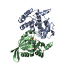 1tx4S S: Starting model for refinement |
|---|---|
| Similar structure data |


-Links
 PDBj
PDBj


- Assembly
| Deposited unit | 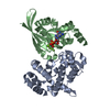
| ||||||||
|---|---|---|---|---|---|---|---|---|---|
| 1 |
| ||||||||
| Unit cell |
|
-Components
-Protein , 2 types, 2 molecules AB
| #1: Protein | Mass: 27444.469 Da / Num. of mol.: 1 Source method: isolated from a genetically manipulated source Source: (gene. exp.) Homo sapiens (human) / Gene: RHOGAP1 / Production host:  Escherichia coli (E. coli) / References: UniProt: Q07960 Escherichia coli (E. coli) / References: UniProt: Q07960 |
|---|---|
| #2: Protein | / H12 Mass: 21766.088 Da / Num. of mol.: 1 Source method: isolated from a genetically manipulated source Source: (gene. exp.) Homo sapiens (human) / Gene: RHOA / Production host: Escherichia coli (E. coli) / References: UniProt: P61586 |
-Non-polymers , 4 types, 377 molecules 


| #3: Chemical | ChemComp-MG /  Mass: 24.305 Da / Num. of mol.: 1 / Source method: obtained synthetically / Formula: Mg Mass: 24.305 Da / Num. of mol.: 1 / Source method: obtained synthetically / Formula: Mg |
|---|---|
| #4: Chemical | ChemComp-GDP / Guanosine diphosphate Type: RNA linking / Mass: 443.201 Da / Num. of mol.: 1 / Source method: obtained synthetically / Formula: C10H15N5O11P2 / Comment: GDP, energy-carrying molecule*YM Type: RNA linking / Mass: 443.201 Da / Num. of mol.: 1 / Source method: obtained synthetically / Formula: C10H15N5O11P2 / Comment: GDP, energy-carrying molecule*YM |
| #5: Chemical | ChemComp-MGF /  Mass: 81.300 Da / Num. of mol.: 1 / Source method: obtained synthetically / Formula: F3Mg Mass: 81.300 Da / Num. of mol.: 1 / Source method: obtained synthetically / Formula: F3Mg |
| #6: Water | ChemComp-HOH / WaterMass: 18.015 Da / Num. of mol.: 374 / Source method: isolated from a natural source / Formula: H2O |
-Experimental details
-Experiment
| Experiment | Method: X-RAY DIFFRACTION / Number of used crystals: 1 |
|---|
- Sample preparation
| Crystal | Density Matthews: 2.2 Å3/Da / Density % sol: 43.63 % | ||||||||||||||||||||||||||||||||||||||||||||||||||||||||||||||||||
|---|---|---|---|---|---|---|---|---|---|---|---|---|---|---|---|---|---|---|---|---|---|---|---|---|---|---|---|---|---|---|---|---|---|---|---|---|---|---|---|---|---|---|---|---|---|---|---|---|---|---|---|---|---|---|---|---|---|---|---|---|---|---|---|---|---|---|---|
| Crystal grow | Temperature: 291 K / Method: vapor diffusion, hanging drop / pH: 6 Details: 18% PEG2KMME, 100mM MES, 10mM MgCl2, 10mM NaF, 2mM deferoxamine, 114mM (NH4)2SO4, pH 6.0, VAPOR DIFFUSION, HANGING DROP, temperature 291K | ||||||||||||||||||||||||||||||||||||||||||||||||||||||||||||||||||
| Crystal grow | *PLUS pH: 7.4 / Details: Rittinger, K., (1997) Nature, 389, 758. | ||||||||||||||||||||||||||||||||||||||||||||||||||||||||||||||||||
| Components of the solutions | *PLUS
|
-Data collection
| Diffraction | Mean temperature: 100 K |
|---|---|
| Diffraction source | Source: SYNCHROTRON / Site: ESRF  / Beamline: BM14 / Wavelength: 1 Å / Beamline: BM14 / Wavelength: 1 Å |
| Radiation | Protocol: SINGLE WAVELENGTH / Monochromatic (M) / Laue (L): M / Scattering type: x-ray |
| Radiation wavelength | Wavelength: 1 Å / Relative weight: 1 |
| Reflection | Resolution: 1.8→15 Å / Num. all: 40798 / Num. obs: 40618 / Observed criterion σ(F): 2 / Observed criterion σ(I): 2 / Redundancy: 5.4 % / Rsym value: 0.083 / Net I/σ(I): 17.5 |
| Reflection shell | Resolution: 1.8→1.86 Å / Mean I/σ(I) obs: 3.7 / Rsym value: 0.401 / % possible all: 99.6 |
| Reflection | *PLUS Lowest resolution: 15 Å / % possible obs: 99.6 % / Num. measured all: 218823 / Rmerge(I) obs: 0.083 |
| Reflection shell | *PLUS % possible obs: 99.9 % / Rmerge(I) obs: 0.401 |
- Processing
| Software |
| |||||||||||||||||||||||||||||||||||||||||||||||||||||||||||||||||||||||||||
|---|---|---|---|---|---|---|---|---|---|---|---|---|---|---|---|---|---|---|---|---|---|---|---|---|---|---|---|---|---|---|---|---|---|---|---|---|---|---|---|---|---|---|---|---|---|---|---|---|---|---|---|---|---|---|---|---|---|---|---|---|---|---|---|---|---|---|---|---|---|---|---|---|---|---|---|---|
| Refinement | Method to determine structure: MOLECULAR REPLACEMENT Starting model: 1TX4 Resolution: 1.8→15 Å / Cor.coef. Fo:Fc: 0.953 / Cor.coef. Fo:Fc free: 0.93 / SU B: 2.434 / SU ML: 0.077 / Cross valid method: THROUGHOUT / ESU R: 0.126 / ESU R Free: 0.119
| |||||||||||||||||||||||||||||||||||||||||||||||||||||||||||||||||||||||||||
| Solvent computation | Ion probe radii: 0.8 Å / Shrinkage radii: 0.8 Å / VDW probe radii: 1.4 Å / Solvent model: BABINET MODEL WITH MASK | |||||||||||||||||||||||||||||||||||||||||||||||||||||||||||||||||||||||||||
| Displacement parameters | Biso mean: 22.779 Å2
| |||||||||||||||||||||||||||||||||||||||||||||||||||||||||||||||||||||||||||
| Refinement step | Cycle: LAST / Resolution: 1.8→15 Å
| |||||||||||||||||||||||||||||||||||||||||||||||||||||||||||||||||||||||||||
| Refine LS restraints |
| |||||||||||||||||||||||||||||||||||||||||||||||||||||||||||||||||||||||||||
| LS refinement shell | Resolution: 1.802→1.848 Å / Total num. of bins used: 20 /
| |||||||||||||||||||||||||||||||||||||||||||||||||||||||||||||||||||||||||||
| Software | *PLUS Version: 5 / Classification: refinement | |||||||||||||||||||||||||||||||||||||||||||||||||||||||||||||||||||||||||||
| Refinement | *PLUS Highest resolution: 1.8 Å / Lowest resolution: 15 Å / Rfactor Rfree: 0.22 / Rfactor Rwork: 0.183 | |||||||||||||||||||||||||||||||||||||||||||||||||||||||||||||||||||||||||||
| Solvent computation | *PLUS | |||||||||||||||||||||||||||||||||||||||||||||||||||||||||||||||||||||||||||
| Displacement parameters | *PLUS | |||||||||||||||||||||||||||||||||||||||||||||||||||||||||||||||||||||||||||
| Refine LS restraints | *PLUS
| |||||||||||||||||||||||||||||||||||||||||||||||||||||||||||||||||||||||||||
| LS refinement shell | *PLUS Rfactor Rfree: 0.244 / Rfactor Rwork: 0.205 |
