 Movie
Movie Controller


+ Open data

- Basic information
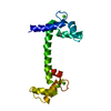
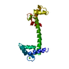
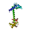
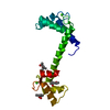
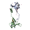
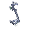
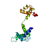
| Entry | Database: PDB / ID: 1ooj | ||||||
|---|---|---|---|---|---|---|---|
| Title | Structural genomics of Caenorhabditis elegans : Calmodulin | ||||||
 Components Components | Calmodulin CMD-1 | ||||||
 Keywords Keywords | METAL BINDING PROTEIN /  Structural genomics / Calmodulin / PSI / Protein Structure Initiative / Southeast Collaboratory for Structural Genomics / SECSG Structural genomics / Calmodulin / PSI / Protein Structure Initiative / Southeast Collaboratory for Structural Genomics / SECSG | ||||||
| Function / homology |  Function and homology information Function and homology informationestablishment of meiotic spindle orientation / Neutrophil degranulation / calcineurin complex / apoptotic cell clearance / embryo development ending in birth or egg hatching / positive chemotaxis / enzyme regulator activity / cell periphery / mitotic spindle / regulation of protein localization ...establishment of meiotic spindle orientation / Neutrophil degranulation / calcineurin complex / apoptotic cell clearance / embryo development ending in birth or egg hatching / positive chemotaxis / enzyme regulator activity / cell periphery / mitotic spindle / regulation of protein localization / cell migration / regulation of apoptotic process / nuclear membrane / regulation of cell cycle / negative regulation of gene expression / centrosome / calcium ion bindingSimilarity search - Function | ||||||
| Biological species |  Caenorhabditis elegans (invertebrata) Caenorhabditis elegans (invertebrata) | ||||||
| Method | X-RAY DIFFRACTION / SYNCHROTRON / MOLECULAR REPLACEMENT / Resolution: 2.11 Å | ||||||
 Authors Authors | Symersky, J. / Lin, G. / Li, S. / Qiu, S. / Luan, C.-H. / Luo, D. / Tsao, J. / Carson, M. / DeLucas, L. / Luo, M. / Southeast Collaboratory for Structural Genomics (SECSG) | ||||||
 Citation Citation | Journal: Proteins / Year: 2003 Title: Structural genomics of caenorhabditis elegans: crystal structure of calmodulin. Authors: Symersky, J. / Lin, G. / Li, S. / Qiu, S. / Carson, M. / Schormann, N. / Luo, M. | ||||||
| History |
|
- Structure visualization
| Structure viewer | Molecule: MolmilJmol/JSmol |
|---|
- Downloads & links
-Download
| PDBx/mmCIF format | 1ooj.cif.gz | 45.6 KB | Display | PDBx/mmCIF format |
|---|---|---|---|---|
| PDB format | pdb1ooj.ent.gz | 30.8 KB | Display | PDB format |
| PDBx/mmJSON format | 1ooj.json.gz | Tree view | PDBx/mmJSON format | |
| Others |  Other downloads Other downloads |
-Validation report
| Arichive directory | https://data.pdbj.org/pub/pdb/validation_reports/oo/1oojftp://data.pdbj.org/pub/pdb/validation_reports/oo/1ooj | HTTPS FTP |
|---|
-Related structure data
| Related structure data |  4clnS S: Starting model for refinement |
|---|---|
| Similar structure data | |
| Other databases |


-Links
 PDBj
PDBj
- Assembly
| Deposited unit | 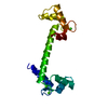
| ||||||||
|---|---|---|---|---|---|---|---|---|---|
| 1 |
| ||||||||
| Unit cell |
|
-Components
| #1: Protein | Mass: 16839.545 Da / Num. of mol.: 1 Source method: isolated from a genetically manipulated source Source: (gene. exp.) Caenorhabditis elegans (invertebrata) / Plasmid: pDEST 17.1 / Production host:  Escherichia coli (E. coli) / References: UniProt: O16305 Escherichia coli (E. coli) / References: UniProt: O16305 | ||
|---|---|---|---|
| #2: Chemical | ChemComp-CA /   Mass: 40.078 Da / Num. of mol.: 4 / Source method: obtained synthetically / Formula: Ca Mass: 40.078 Da / Num. of mol.: 4 / Source method: obtained synthetically / Formula: Ca#3: Water | ChemComp-HOH / | Water Mass: 18.015 Da / Num. of mol.: 91 / Source method: isolated from a natural source / Formula: H2O Mass: 18.015 Da / Num. of mol.: 91 / Source method: isolated from a natural source / Formula: H2O |
-Experimental details
-Experiment
| Experiment | Method: X-RAY DIFFRACTION / Number of used crystals: 1 |
|---|
- Sample preparation
| Crystal | Density Matthews: 2.07 Å3/Da / Density % sol: 40.05 % | ||||||||||||||||||||||||||||||||||||||||||||||||||||||||||||||||||||||
|---|---|---|---|---|---|---|---|---|---|---|---|---|---|---|---|---|---|---|---|---|---|---|---|---|---|---|---|---|---|---|---|---|---|---|---|---|---|---|---|---|---|---|---|---|---|---|---|---|---|---|---|---|---|---|---|---|---|---|---|---|---|---|---|---|---|---|---|---|---|---|---|
| Crystal grow | Temperature: 295 K / Method: vapor diffusion, hanging drop / pH: 5.7 Details: PH 5.7, VAPOR DIFFUSION, HANGING DROP, TEMPERATURE 295.0K, RESERVOIR: MPD, PEG3350, DIOXANE, 5 MM CACL2, 50 MM CITRATE, PH 5.7, PROTEIN STOCK: 5 MG/ML IN 5 MM CACL2, 5 MM CACODYLATE, PH 6, ...Details: PH 5.7, VAPOR DIFFUSION, HANGING DROP, TEMPERATURE 295.0K, RESERVOIR: MPD, PEG3350, DIOXANE, 5 MM CACL2, 50 MM CITRATE, PH 5.7, PROTEIN STOCK: 5 MG/ML IN 5 MM CACL2, 5 MM CACODYLATE, PH 6, DROPS: 5 MICROLITERS OF PROTEIN STOCK SOLUTION, 5 MICROLITERS OF RESERVOIR. | ||||||||||||||||||||||||||||||||||||||||||||||||||||||||||||||||||||||
| Crystal grow | *PLUS pH: 6 | ||||||||||||||||||||||||||||||||||||||||||||||||||||||||||||||||||||||
| Components of the solutions | *PLUS
|
-Data collection
| Diffraction | Mean temperature: 100 K |
|---|---|
| Diffraction source | Source: SYNCHROTRON / Site: APS  / Beamline: 14-BM-D / Wavelength: 1.107 Å / Beamline: 14-BM-D / Wavelength: 1.107 Å |
| Detector | Type: ADSC QUANTUM 4 / Detector: CCD / Date: Aug 8, 2002 |
| Radiation | Protocol: SINGLE WAVELENGTH / Monochromatic (M) / Laue (L): M / Scattering type: x-ray |
| Radiation wavelength | Wavelength: 1.107 Å / Relative weight: 1 |
| Reflection | Resolution: 2.11→50 Å / Num. all: 8241 / Num. obs: 8241 / % possible obs: 99.7 % / Observed criterion σ(F): 0 / Observed criterion σ(I): 0 / Redundancy: 3.2 % / Biso Wilson estimate: 26.6 Å2 / Rsym value: 0.071 / Net I/σ(I): 12 |
| Reflection shell | Resolution: 2.11→2.19 Å / Redundancy: 3.2 % / Mean I/σ(I) obs: 9.3 / Num. unique all: 839 / Rsym value: 0.202 / % possible all: 99.8 |
| Reflection | *PLUS Lowest resolution: 30 Å / Num. measured all: 29757 / Rmerge(I) obs: 0.071 |
| Reflection shell | *PLUS % possible obs: 99.8 % / Rmerge(I) obs: 0.202 |
- Processing
| Software |
| ||||||||||||||||||||||||||||||||||||
|---|---|---|---|---|---|---|---|---|---|---|---|---|---|---|---|---|---|---|---|---|---|---|---|---|---|---|---|---|---|---|---|---|---|---|---|---|---|
| Refinement | Method to determine structure: MOLECULAR REPLACEMENT Starting model: PDB ENTRY 4CLN Resolution: 2.11→30 Å / Rfactor Rfree error: 0.014 / Data cutoff high absF: 10000 / Data cutoff high rms absF: 10000 / Isotropic thermal model: restrained / Cross valid method: THROUGHOUT / σ(F): 4 / Stereochemistry target values: Engh & Huber
| ||||||||||||||||||||||||||||||||||||
| Solvent computation | Solvent model: flat model / Bsol: 68.53 Å2 / ksol: 0.4 e/Å3 | ||||||||||||||||||||||||||||||||||||
| Displacement parameters | Biso mean: 33.7 Å2 | ||||||||||||||||||||||||||||||||||||
| Refine analyze |
| ||||||||||||||||||||||||||||||||||||
| Refinement step | Cycle: LAST / Resolution: 2.11→30 Å
| ||||||||||||||||||||||||||||||||||||
| Refine LS restraints |
| ||||||||||||||||||||||||||||||||||||
| LS refinement shell | Resolution: 2.11→2.19 Å / Rfactor Rfree error: 0.046 / Total num. of bins used: 10
| ||||||||||||||||||||||||||||||||||||
| Xplor file |
| ||||||||||||||||||||||||||||||||||||
| Refinement | *PLUS Lowest resolution: 30 Å / % reflection Rfree: 5 % | ||||||||||||||||||||||||||||||||||||
| Solvent computation | *PLUS | ||||||||||||||||||||||||||||||||||||
| Displacement parameters | *PLUS | ||||||||||||||||||||||||||||||||||||
| Refine LS restraints | *PLUS
|
