 Movie
Movie Controller

[English] 日本語
 Yorodumi
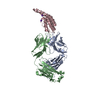
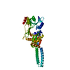
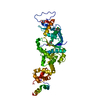
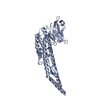
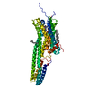
- PDB-1oms: Structure determination by MAD: E.coli Trigger Factor binding at ... -
+ Open data

- Basic information
| Entry | Database: PDB / ID: 1oms | ||||||
|---|---|---|---|---|---|---|---|
| Title | Structure determination by MAD: E.coli Trigger Factor binding at the ribosomal exit tunnel. | ||||||
 Components Components | Trigger Factor | ||||||
 Keywords Keywords |  ISOMERASE / alpha-beta structure ISOMERASE / alpha-beta structure | ||||||
| Function / homology |  Function and homology information Function and homology information'de novo' cotranslational protein folding / stress response to copper ion / protein unfolding / chaperone-mediated protein folding / protein folding chaperone / peptidylprolyl isomerase / peptidyl-prolyl cis-trans isomerase activity / ribosome binding / protein transport / response to heat ...'de novo' cotranslational protein folding / stress response to copper ion / protein unfolding / chaperone-mediated protein folding / protein folding chaperone / peptidylprolyl isomerase / peptidyl-prolyl cis-trans isomerase activity / ribosome binding / protein transport / response to heat / cell cycle / cell division / membrane / identical protein binding / cytosolSimilarity search - Function | ||||||
| Biological species |  Escherichia coli (E. coli) Escherichia coli (E. coli) | ||||||
| Method | X-RAY DIFFRACTION / SYNCHROTRON / MAD / Resolution: 2.3 Å | ||||||
 Authors Authors | Kristensen, O. / Gajhede, M. | ||||||
 Citation Citation | Journal: Structure / Year: 2003 Title: Chaperone binding at the ribosomal exit tunnel. Authors: Kristensen, O. / Gajhede, M. #1: Journal: Nature / Year: 2002Title: L23 protein functions as a chaperone docking site on the ribosome. Authors: Kramer, G. / Rauch, T. / Rist, W. / Vorderwulbecke, S. / Patzelt, H. / Schulze-Specking, A. / Ban, N. / Deuerling, E. / Bukau, B. #2: Journal: J.Mol.Biol. / Year: 2003Title: Localization of the Trigger Factor Binding Site on the Ribosomal 50S Subunit. Authors: Blaha, G. / Wilson, D.N. / Stoller, G. / Fischer, G. / Willumeit, R. / Nierhaus, K.H. #3: Journal: J.Mol.Biol. / Year: 2003Title: Interaction of trigger factor with the ribosome. Authors: Maier, R. / Eckert, B. / Scholz, C. / Lilie, H. / Schmid, F.X. | ||||||
| History |
|
- Structure visualization
| Structure viewer | Molecule: MolmilJmol/JSmol |
|---|
- Downloads & links
-Download
| PDBx/mmCIF format | 1oms.cif.gz | 85.2 KB | Display | PDBx/mmCIF format |
|---|---|---|---|---|
| PDB format | pdb1oms.ent.gz | 71.1 KB | Display | PDB format |
| PDBx/mmJSON format | 1oms.json.gz | Tree view | PDBx/mmJSON format | |
| Others |  Other downloads Other downloads |
-Validation report
| Arichive directory | https://data.pdbj.org/pub/pdb/validation_reports/om/1omsftp://data.pdbj.org/pub/pdb/validation_reports/om/1oms | HTTPS FTP |
|---|
-Related structure data


-Links
 PDBj
PDBj

- Assembly
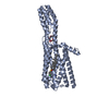
| Deposited unit | 
| |||||||||||||||||||||
|---|---|---|---|---|---|---|---|---|---|---|---|---|---|---|---|---|---|---|---|---|---|---|
| 1 |
| |||||||||||||||||||||
| Unit cell |
| |||||||||||||||||||||
| Components on special symmetry positions |
|
-Components
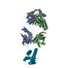
-Protein , 1 types, 3 molecules ABC
| #1: Protein | Mass: 13504.907 Da / Num. of mol.: 3 / Fragment: Ribosome binding domain Source method: isolated from a genetically manipulated source Source: (gene. exp.) Escherichia coli (E. coli) / Gene: tig / Plasmid: pET28a / Production host: Escherichia coli (E. coli) / Strain (production host): BL21(DE3)pLysS / References: UniProt: P0A850, peptidylprolyl isomerase |
|---|
-Non-polymers , 5 types, 234 molecules 


| #2: Chemical | ChemComp-SO4 / Sulfate Mass: 96.063 Da / Num. of mol.: 13 / Source method: obtained synthetically / Formula: SO4 Mass: 96.063 Da / Num. of mol.: 13 / Source method: obtained synthetically / Formula: SO4#3: Chemical | ChemComp-PG4 / Polyethylene glycol Mass: 194.226 Da / Num. of mol.: 5 / Source method: obtained synthetically / Formula: C8H18O5 / Comment: precipitant*YM Mass: 194.226 Da / Num. of mol.: 5 / Source method: obtained synthetically / Formula: C8H18O5 / Comment: precipitant*YM#4: Chemical | ChemComp-SO2 / | Sulfur dioxide Mass: 64.064 Da / Num. of mol.: 1 / Source method: obtained synthetically / Formula: O2S Mass: 64.064 Da / Num. of mol.: 1 / Source method: obtained synthetically / Formula: O2S#5: Chemical | ChemComp-GOL / | Glycerol Mass: 92.094 Da / Num. of mol.: 1 / Source method: obtained synthetically / Formula: C3H8O3 Mass: 92.094 Da / Num. of mol.: 1 / Source method: obtained synthetically / Formula: C3H8O3#6: Water | ChemComp-HOH / | WaterMass: 18.015 Da / Num. of mol.: 214 / Source method: isolated from a natural source / Formula: H2O |
|---|
-Experimental details
-Experiment
| Experiment | Method: X-RAY DIFFRACTION / Number of used crystals: 1 |
|---|
- Sample preparation
| Crystal | Density Matthews: 2.16 Å3/Da / Density % sol: 42.62 % | ||||||||||||||||||||||||||||||
|---|---|---|---|---|---|---|---|---|---|---|---|---|---|---|---|---|---|---|---|---|---|---|---|---|---|---|---|---|---|---|---|
| Crystal grow | Temperature: 279 K / Method: vapor diffusion, hanging drop / pH: 4.6 Details: PEG 2000 MME, PEG 400, ammonium sulfate, sodium acetate, tris, pH 4.6, VAPOR DIFFUSION, HANGING DROP, temperature 279K | ||||||||||||||||||||||||||||||
| Crystal grow | *PLUS Temperature: 6 ℃ / Method: vapor diffusion, hanging drop | ||||||||||||||||||||||||||||||
| Components of the solutions | *PLUS
|
-Data collection
| Diffraction | Mean temperature: 100 K | |||||||||
|---|---|---|---|---|---|---|---|---|---|---|
| Diffraction source | Source: SYNCHROTRON / Site: EMBL/DESY, Hamburg  / Beamline: BW7A / Wavelength: 0.9797, 0.9404 / Beamline: BW7A / Wavelength: 0.9797, 0.9404 | |||||||||
| Detector | Type: MARRESEARCH / Detector: CCD / Date: Nov 28, 2002 | |||||||||
| Radiation | Monochromator: sagittal-focusing double crystal monochromator in combination with a vertical focusing mirror. Protocol: MAD / Monochromatic (M) / Laue (L): M / Scattering type: x-ray | |||||||||
| Radiation wavelength |
| |||||||||
| Reflection | Resolution: 2.3→30 Å / Num. obs: 17023 / % possible obs: 99.6 % / Observed criterion σ(F): 0 / Observed criterion σ(I): 0 / Redundancy: 7.8 % / Biso Wilson estimate: 22.6 Å2 / Rmerge(I) obs: 0.059 / Rsym value: 0.059 / Net I/σ(I): 33.3 | |||||||||
| Reflection shell | Resolution: 2.3→2.42 Å / Redundancy: 7.8 % / Rmerge(I) obs: 0.33 / Mean I/σ(I) obs: 6.6 / Num. unique all: 2432 / Rsym value: 0.33 / % possible all: 100 | |||||||||
| Reflection | *PLUS Lowest resolution: 30 Å / Num. obs: 31810 / Redundancy: 4.2 % | |||||||||
| Reflection shell | *PLUS Highest resolution: 2.3 Å / % possible obs: 100 % / Redundancy: 4.1 % / Rmerge(I) obs: 0.33 |
- Processing
| Software |
| ||||||||||||||||||||||||||||||||||||
|---|---|---|---|---|---|---|---|---|---|---|---|---|---|---|---|---|---|---|---|---|---|---|---|---|---|---|---|---|---|---|---|---|---|---|---|---|---|
| Refinement | Method to determine structure: MAD / Resolution: 2.3→29.9 Å / Rfactor Rfree error: 0.005 / Data cutoff high absF: 5499572.78 / Data cutoff low absF: 0 / Isotropic thermal model: RESTRAINED / Cross valid method: THROUGHOUT / σ(F): 0 / Stereochemistry target values: Engh & Huber Details: Refinement was based on the "mlhl" target function. Experimental phases and strong NCS restraints were used throughout. Used all data in the ultimate brief refinement cycle against a ...Details: Refinement was based on the "mlhl" target function. Experimental phases and strong NCS restraints were used throughout. Used all data in the ultimate brief refinement cycle against a standard crystallographic target: R-factor(all).
| ||||||||||||||||||||||||||||||||||||
| Solvent computation | Solvent model: FLAT MODEL / Bsol: 39.9718 Å2 / ksol: 0.353647 e/Å3 | ||||||||||||||||||||||||||||||||||||
| Displacement parameters | Biso mean: 35.8 Å2
| ||||||||||||||||||||||||||||||||||||
| Refine analyze |
| ||||||||||||||||||||||||||||||||||||
| Refinement step | Cycle: LAST / Resolution: 2.3→29.9 Å
| ||||||||||||||||||||||||||||||||||||
| Refine LS restraints |
| ||||||||||||||||||||||||||||||||||||
| LS refinement shell | Resolution: 2.3→2.44 Å / Rfactor Rfree error: 0.018 / Total num. of bins used: 6
| ||||||||||||||||||||||||||||||||||||
| Xplor file |
| ||||||||||||||||||||||||||||||||||||
| Refinement | *PLUS % reflection Rfree: 5 % | ||||||||||||||||||||||||||||||||||||
| Solvent computation | *PLUS | ||||||||||||||||||||||||||||||||||||
| Displacement parameters | *PLUS | ||||||||||||||||||||||||||||||||||||
| Refine LS restraints | *PLUS
|
