 Movie
Movie Controller

[English] 日本語
 Yorodumi
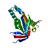
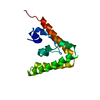
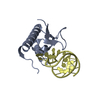
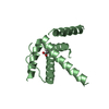
- PDB-1oj8: Novel and retro Binding Modes in Cytotoxic Ribonucleases from Ran... -
+ Open data

- Basic information
| Entry | Database: PDB / ID: 1oj8 | |||||||||
|---|---|---|---|---|---|---|---|---|---|---|
| Title | Novel and retro Binding Modes in Cytotoxic Ribonucleases from Rana catesbeiana of Two Crystal Structures Complexed with d(ApCpGpA) and (2',5'CpG) | |||||||||
 Components Components |
| |||||||||
 Keywords Keywords |  HYDROLASE / CYTOTOXIC RIBONUCLEASES / ANTI-TUMOR ACTIVITY / SIALIC BINDING AND NUCLEOTIDE BINDING HYDROLASE / CYTOTOXIC RIBONUCLEASES / ANTI-TUMOR ACTIVITY / SIALIC BINDING AND NUCLEOTIDE BINDING | |||||||||
| Function / homology |  Function and homology information Function and homology informationP-30 Protein / Ribonuclease A-like domain / Ribonuclease A, active site / Ribonuclease A-domain / Ribonuclease A-like domain superfamily / Pancreatic ribonuclease / Pancreatic ribonuclease family signature. / Pancreatic ribonuclease / Roll / Alpha BetaSimilarity search - Domain/homology | |||||||||
| Biological species | RANA CATESBEIANA (American bullfrog) | |||||||||
| Method | X-RAY DIFFRACTION / MOLECULAR REPLACEMENT / Resolution: 1.7 Å | |||||||||
 Authors Authors | Tsai, C.-J. / Liu, J.-H. / Liao, Y.-D. / Chen, L.-Y. / Cheng, P.-T. / Sun, Y.-J. | |||||||||
 Citation Citation | Journal: To be Published Title: Retro and Novel Binding Modes in Cytotoxic Ribonucleases from Rana Catesbeiana of Two Crystal Structures Complexed with (2',5'Cpg) and D(Apcpgpa) Authors: Tsai, C.-J. / Liu, J.-H. / Liao, Y.-D. / Chen, L.-Y. / Cheng, P.-T. / Sun, Y.-J. | |||||||||
| History |
|
- Structure visualization
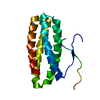
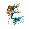
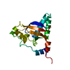
| Structure viewer | Molecule: MolmilJmol/JSmol |
|---|
- Downloads & links
-Download
| PDBx/mmCIF format | 1oj8.cif.gz | 45 KB | Display | PDBx/mmCIF format |
|---|---|---|---|---|
| PDB format | pdb1oj8.ent.gz | 29.6 KB | Display | PDB format |
| PDBx/mmJSON format | 1oj8.json.gz | Tree view | PDBx/mmJSON format | |
| Others |  Other downloads Other downloads |
-Validation report
| Arichive directory | https://data.pdbj.org/pub/pdb/validation_reports/oj/1oj8ftp://data.pdbj.org/pub/pdb/validation_reports/oj/1oj8 | HTTPS FTP |
|---|
-Related structure data
| Related structure data |  1oncS S: Starting model for refinement |
|---|---|
| Similar structure data |


-Links
 PDBj
PDBj

- Assembly
| Deposited unit | 
| ||||||||||||
|---|---|---|---|---|---|---|---|---|---|---|---|---|---|
| 1 |
| ||||||||||||
| Unit cell |
| ||||||||||||
| Components on special symmetry positions |
|
-Components
| #1: Protein | Mass: 12185.388 Da / Num. of mol.: 1 / Source method: isolated from a natural source / Source: (natural) RANA CATESBEIANA (American bullfrog) / Tissue: OCCYTE / References: UniProt: Q9DFY5 | ||
|---|---|---|---|
| #2: DNA chain | Mass: 1199.844 Da / Num. of mol.: 1 / Source method: obtained synthetically | ||
| #3: Chemical | Sulfate  Mass: 96.063 Da / Num. of mol.: 2 / Source method: obtained synthetically / Formula: SO4 Mass: 96.063 Da / Num. of mol.: 2 / Source method: obtained synthetically / Formula: SO4#4: Water | ChemComp-HOH / | Water Mass: 18.015 Da / Num. of mol.: 220 / Source method: isolated from a natural source / Formula: H2O Mass: 18.015 Da / Num. of mol.: 220 / Source method: isolated from a natural source / Formula: H2O |
-Experimental details
-Experiment
| Experiment | Method: X-RAY DIFFRACTION / Number of used crystals: 1 |
|---|
- Sample preparation
| Crystal | Density Matthews: 2.59 Å3/Da / Density % sol: 53 % |
|---|---|
| Crystal grow | pH: 7 / Details: 3.3 M AMMONIUM SULFATE,2% PEG 400, pH 7.0 |
-Data collection
| Diffraction | Mean temperature: 100 K |
|---|---|
| Diffraction source | Source: ROTATING ANODE / Type: RIGAKU RU300 / Wavelength: 1.54 |
| Detector | Type: RIGAKU IMAGE PLATE / Detector: IMAGE PLATE / Details: CONFOCAL |
| Radiation | Protocol: SINGLE WAVELENGTH / Monochromatic (M) / Laue (L): M / Scattering type: x-ray |
| Radiation wavelength | Wavelength: 1.54 Å / Relative weight: 1 |
| Reflection | Resolution: 1.7→20 Å / Num. obs: 12965 / % possible obs: 90.5 % / Observed criterion σ(I): 2 / Redundancy: 7.32 % / Biso Wilson estimate: 20.7 Å2 / Rmerge(I) obs: 0.044 / Net I/σ(I): 35.5 |
| Reflection shell | Resolution: 1.7→1.76 Å / Redundancy: 7.3 % / Rmerge(I) obs: 0.43 / Mean I/σ(I) obs: 4.8 / % possible all: 97.5 |
- Processing
| Software |
| ||||||||||||||||||||||||||||||||||||||||||||||||||||||||||||||||||||||||||||||||
|---|---|---|---|---|---|---|---|---|---|---|---|---|---|---|---|---|---|---|---|---|---|---|---|---|---|---|---|---|---|---|---|---|---|---|---|---|---|---|---|---|---|---|---|---|---|---|---|---|---|---|---|---|---|---|---|---|---|---|---|---|---|---|---|---|---|---|---|---|---|---|---|---|---|---|---|---|---|---|---|---|---|
| Refinement | Method to determine structure: MOLECULAR REPLACEMENT Starting model: PDB ENTRY 1ONC Resolution: 1.7→20 Å / Rfactor Rfree error: 0.008 / Isotropic thermal model: RESTRAINED / Cross valid method: THROUGHOUT / σ(F): 2
| ||||||||||||||||||||||||||||||||||||||||||||||||||||||||||||||||||||||||||||||||
| Solvent computation | Solvent model: FLAT MODEL / Bsol: 50.9 Å2 / ksol: 0.38 e/Å3 | ||||||||||||||||||||||||||||||||||||||||||||||||||||||||||||||||||||||||||||||||
| Displacement parameters | Biso mean: 29.6 Å2
| ||||||||||||||||||||||||||||||||||||||||||||||||||||||||||||||||||||||||||||||||
| Refine analyze |
| ||||||||||||||||||||||||||||||||||||||||||||||||||||||||||||||||||||||||||||||||
| Refinement step | Cycle: LAST / Resolution: 1.7→20 Å
| ||||||||||||||||||||||||||||||||||||||||||||||||||||||||||||||||||||||||||||||||
| Refine LS restraints |
| ||||||||||||||||||||||||||||||||||||||||||||||||||||||||||||||||||||||||||||||||
| LS refinement shell | Resolution: 1.7→1.81 Å / Rfactor Rfree error: 0.026 / Total num. of bins used: 6
| ||||||||||||||||||||||||||||||||||||||||||||||||||||||||||||||||||||||||||||||||
| Xplor file |
|
