 Movie
Movie Controller


+ Open data

- Basic information
| Entry | Database: PDB / ID: 1ohw | ||||||
|---|---|---|---|---|---|---|---|
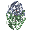
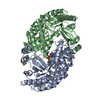
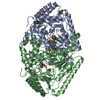
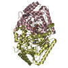
| Title | 4-AMINOBUTYRATE-AMINOTRANSFERASE inactivated by gamma-vinyl GABA | ||||||
 Components Components | 4-AMINOBUTYRATE AMINOTRANSFERASE | ||||||
 Keywords Keywords |  TRANSFERASE / PLP-DEPENDENT ENZYME / AMINOTRANSFERASE / 4- AMINOBUTYRIC ACID / ANTIEPILEPTIC DRUG TARGET / VIGABATRIN PYRIDOXAL PHOSPHATE / NEUROTRANSMITTER DEGRADATION / MITOCHONDRION / TRANSIT PEPTIDE TRANSFERASE / PLP-DEPENDENT ENZYME / AMINOTRANSFERASE / 4- AMINOBUTYRIC ACID / ANTIEPILEPTIC DRUG TARGET / VIGABATRIN PYRIDOXAL PHOSPHATE / NEUROTRANSMITTER DEGRADATION / MITOCHONDRION / TRANSIT PEPTIDE | ||||||
| Function / homology |  Function and homology information(S)-3-amino-2-methylpropionate transaminase / 4-aminobutyrate transaminase complex / succinate-semialdehyde dehydrogenase binding / (S)-3-amino-2-methylpropionate transaminase activity / Degradation of GABA / 4-aminobutyrate-2-oxoglutarate transaminase / 4-aminobutyrate:2-oxoglutarate transaminase activity / 4-aminobutyrate transaminase activity / gamma-aminobutyric acid catabolic process / behavioral response to cocaine ...(S)-3-amino-2-methylpropionate transaminase / 4-aminobutyrate transaminase complex / succinate-semialdehyde dehydrogenase binding / (S)-3-amino-2-methylpropionate transaminase activity / Degradation of GABA / 4-aminobutyrate-2-oxoglutarate transaminase / 4-aminobutyrate:2-oxoglutarate transaminase activity / 4-aminobutyrate transaminase activity / gamma-aminobutyric acid catabolic process / behavioral response to cocaine / 2 iron, 2 sulfur cluster binding / pyridoxal phosphate binding / mitochondrial matrix / protein homodimerization activity / mitochondrion / identical protein binding / metal ion binding / cytosol Function and homology information(S)-3-amino-2-methylpropionate transaminase / 4-aminobutyrate transaminase complex / succinate-semialdehyde dehydrogenase binding / (S)-3-amino-2-methylpropionate transaminase activity / Degradation of GABA / 4-aminobutyrate-2-oxoglutarate transaminase / 4-aminobutyrate:2-oxoglutarate transaminase activity / 4-aminobutyrate transaminase activity / gamma-aminobutyric acid catabolic process / behavioral response to cocaine ...(S)-3-amino-2-methylpropionate transaminase / 4-aminobutyrate transaminase complex / succinate-semialdehyde dehydrogenase binding / (S)-3-amino-2-methylpropionate transaminase activity / Degradation of GABA / 4-aminobutyrate-2-oxoglutarate transaminase / 4-aminobutyrate:2-oxoglutarate transaminase activity / 4-aminobutyrate transaminase activity / gamma-aminobutyric acid catabolic process / behavioral response to cocaine / 2 iron, 2 sulfur cluster binding / pyridoxal phosphate binding / mitochondrial matrix / protein homodimerization activity / mitochondrion / identical protein binding / metal ion binding / cytosolSimilarity search - Function | ||||||
| Biological species |  SUS SCROFA (pig) SUS SCROFA (pig) | ||||||
| Method | X-RAY DIFFRACTION / SYNCHROTRON / OTHER / Resolution: 2.3 Å | ||||||
 Authors Authors | Storici, P. / Schirmer, T. | ||||||
 Citation Citation | Journal: J.Biol.Chem. / Year: 2004 Title: Structures of {Gamma}-Aminobutyric Acid (Gaba) Aminotransferase, a Pyridoxal 5'-Phosphate, and [2Fe-2S] Cluster-Containing Enzyme, Complexed with {Gamma}-Ethynyl-Gaba and with the Antiepilepsy Drug Vigabatrin Authors: Storici, P. / De Biase, D. / Bossa, F. / Bruno, S. / Mozzarelli, A. / Peneff, C. / Silverman, R. / Schirmer, T. #1: Journal: Biochemistry / Year: 1999 Title: Crystal Structure of Gaba-Aminotransferase, a Target for Antiepileptic Drug Therapy Authors: Storici, P. / Capitani, G. / De Biase, D. / Moser, M. / John, R.A. / Jansonius, J.N. / Schirmer, T. | ||||||
| History |
|
- Structure visualization
| Structure viewer | Molecule: MolmilJmol/JSmol |
|---|
- Downloads & links
-Download
| PDBx/mmCIF format | 1ohw.cif.gz | 363.6 KB | Display | PDBx/mmCIF format |
|---|---|---|---|---|
| PDB format | pdb1ohw.ent.gz | 304.9 KB | Display | PDB format |
| PDBx/mmJSON format | 1ohw.json.gz | Tree view | PDBx/mmJSON format | |
| Others |  Other downloads Other downloads |
-Validation report
| Arichive directory | https://data.pdbj.org/pub/pdb/validation_reports/oh/1ohwftp://data.pdbj.org/pub/pdb/validation_reports/oh/1ohw | HTTPS FTP |
|---|
-Related structure data


-Links
 PDBj
PDBj


- Assembly
| Deposited unit | 
| |||||||||||||||||||||||||||||||||||||||||||||||||||||||||||||||||||||||||||||||||||||||||||||||||||||||||||||||||||||||||||||||||||||||||||||||||||||||||||||||||||||||||||||||||||||||||||||||||||||||||||||||||||||||||||||||||||||
|---|---|---|---|---|---|---|---|---|---|---|---|---|---|---|---|---|---|---|---|---|---|---|---|---|---|---|---|---|---|---|---|---|---|---|---|---|---|---|---|---|---|---|---|---|---|---|---|---|---|---|---|---|---|---|---|---|---|---|---|---|---|---|---|---|---|---|---|---|---|---|---|---|---|---|---|---|---|---|---|---|---|---|---|---|---|---|---|---|---|---|---|---|---|---|---|---|---|---|---|---|---|---|---|---|---|---|---|---|---|---|---|---|---|---|---|---|---|---|---|---|---|---|---|---|---|---|---|---|---|---|---|---|---|---|---|---|---|---|---|---|---|---|---|---|---|---|---|---|---|---|---|---|---|---|---|---|---|---|---|---|---|---|---|---|---|---|---|---|---|---|---|---|---|---|---|---|---|---|---|---|---|---|---|---|---|---|---|---|---|---|---|---|---|---|---|---|---|---|---|---|---|---|---|---|---|---|---|---|---|---|---|---|---|---|---|---|---|---|---|---|---|---|---|---|---|---|---|---|---|---|
| 1 | 
| |||||||||||||||||||||||||||||||||||||||||||||||||||||||||||||||||||||||||||||||||||||||||||||||||||||||||||||||||||||||||||||||||||||||||||||||||||||||||||||||||||||||||||||||||||||||||||||||||||||||||||||||||||||||||||||||||||||
| 2 | 
| |||||||||||||||||||||||||||||||||||||||||||||||||||||||||||||||||||||||||||||||||||||||||||||||||||||||||||||||||||||||||||||||||||||||||||||||||||||||||||||||||||||||||||||||||||||||||||||||||||||||||||||||||||||||||||||||||||||
| Unit cell |
| |||||||||||||||||||||||||||||||||||||||||||||||||||||||||||||||||||||||||||||||||||||||||||||||||||||||||||||||||||||||||||||||||||||||||||||||||||||||||||||||||||||||||||||||||||||||||||||||||||||||||||||||||||||||||||||||||||||
| Noncrystallographic symmetry (NCS) | NCS domain:
NCS domain segments:
NCS oper:
|
-Components
| #1: Protein | Mass: 53368.996 Da / Num. of mol.: 4 / Source method: isolated from a natural source Details: AFTER REACTION WITH 4-AMINO-5-HEXENOIC ACID (GAMMA-VINYL GABA, VIGABATRIN) THE FRAGMENT VIG HAS BEEN GENERATED WHICH IS COVALENTLY ATTACHED VIA A DOUBLE BOND TO C4A OF PLP AND VIA A SINGLE ...Details: AFTER REACTION WITH 4-AMINO-5-HEXENOIC ACID (GAMMA-VINYL GABA, VIGABATRIN) THE FRAGMENT VIG HAS BEEN GENERATED WHICH IS COVALENTLY ATTACHED VIA A DOUBLE BOND TO C4A OF PLP AND VIA A SINGLE BOND TO NZ OF THE ACTIVE SITE LYS 329 Source: (natural) SUS SCROFA (pig) / Organ: LIVERReferences: UniProt: P80147, 4-aminobutyrate-2-oxoglutarate transaminase #2: Chemical | ChemComp-PLP / Pyridoxal phosphate  Mass: 247.142 Da / Num. of mol.: 4 / Source method: obtained synthetically / Formula: C8H10NO6P Mass: 247.142 Da / Num. of mol.: 4 / Source method: obtained synthetically / Formula: C8H10NO6P#3: Chemical | ChemComp-VIG /   Mass: 131.173 Da / Num. of mol.: 4 / Source method: obtained synthetically / Formula: C6H13NO2 Mass: 131.173 Da / Num. of mol.: 4 / Source method: obtained synthetically / Formula: C6H13NO2#4: Chemical | Iron–sulfur cluster  Mass: 175.820 Da / Num. of mol.: 2 / Source method: obtained synthetically / Formula: Fe2S2 Mass: 175.820 Da / Num. of mol.: 2 / Source method: obtained synthetically / Formula: Fe2S2#5: Water | ChemComp-HOH / | Water Mass: 18.015 Da / Num. of mol.: 716 / Source method: isolated from a natural source / Formula: H2O Mass: 18.015 Da / Num. of mol.: 716 / Source method: isolated from a natural source / Formula: H2OCompound details | CATALYTIC ACTIVITY: 4-AMINOBUTAN | |
|---|
-Experimental details
-Experiment
| Experiment | Method: X-RAY DIFFRACTION / Number of used crystals: 1 |
|---|
- Sample preparation
| Crystal | Density Matthews: 2.5 Å3/Da / Density % sol: 50.79 % | ||||||||||||||||||||||||||||||||||||||||||
|---|---|---|---|---|---|---|---|---|---|---|---|---|---|---|---|---|---|---|---|---|---|---|---|---|---|---|---|---|---|---|---|---|---|---|---|---|---|---|---|---|---|---|---|
| Crystal grow | pH: 5.7 / Details: pH 5.70 | ||||||||||||||||||||||||||||||||||||||||||
| Crystal grow | *PLUS pH: 5.4 / Method: vapor diffusion, sitting drop | ||||||||||||||||||||||||||||||||||||||||||
| Components of the solutions | *PLUS
|
-Data collection
| Diffraction | Mean temperature: 100 K |
|---|---|
| Diffraction source | Source: SYNCHROTRON / Site: ELETTRA  / Beamline: 5.2R / Wavelength: 0.984 / Beamline: 5.2R / Wavelength: 0.984 |
| Detector | Type: MARRESEARCH / Detector: IMAGE PLATE |
| Radiation | Protocol: SINGLE WAVELENGTH / Monochromatic (M) / Laue (L): M / Scattering type: x-ray |
| Radiation wavelength | Wavelength: 0.984 Å / Relative weight: 1 |
| Reflection | Resolution: 2.3→30 Å / Num. obs: 88767 / % possible obs: 96.1 % / Redundancy: 2.22 % / Rmerge(I) obs: 0.073 / Net I/σ(I): 6.6779 |
| Reflection shell | Resolution: 2.3→2.42 Å / Redundancy: 2.18 % / Rmerge(I) obs: 0.16 / Mean I/σ(I) obs: 4.14 / % possible all: 96.7 |
| Reflection | *PLUS Highest resolution: 2.3 Å / Lowest resolution: 30 Å / Redundancy: 2.2 % / Rmerge(I) obs: 0.073 |
- Processing
| Software |
| ||||||||||||||||||||||||||||||||||||||||||||||||||||||||||||||||||||||||||||||||||||||||||||||||||||||||||||||||||||||||||||||||||||||||||||||||||||||||||||||||||||||||||||||||||||||
|---|---|---|---|---|---|---|---|---|---|---|---|---|---|---|---|---|---|---|---|---|---|---|---|---|---|---|---|---|---|---|---|---|---|---|---|---|---|---|---|---|---|---|---|---|---|---|---|---|---|---|---|---|---|---|---|---|---|---|---|---|---|---|---|---|---|---|---|---|---|---|---|---|---|---|---|---|---|---|---|---|---|---|---|---|---|---|---|---|---|---|---|---|---|---|---|---|---|---|---|---|---|---|---|---|---|---|---|---|---|---|---|---|---|---|---|---|---|---|---|---|---|---|---|---|---|---|---|---|---|---|---|---|---|---|---|---|---|---|---|---|---|---|---|---|---|---|---|---|---|---|---|---|---|---|---|---|---|---|---|---|---|---|---|---|---|---|---|---|---|---|---|---|---|---|---|---|---|---|---|---|---|---|---|
| Refinement | Method to determine structure: OTHER / Resolution: 2.3→30 Å / Cor.coef. Fo:Fc: 0.938 / Cor.coef. Fo:Fc free: 0.919 / SU B: 6.423 / SU ML: 0.155 / TLS residual ADP flag: LIKELY RESIDUAL / Cross valid method: THROUGHOUT / ESU R: 0.36 / ESU R Free: 0.217 / Stereochemistry target values: MAXIMUM LIKELIHOOD / Details: HYDROGENS HAVE BEEN ADDED IN THE RIDING POSITIONS
| ||||||||||||||||||||||||||||||||||||||||||||||||||||||||||||||||||||||||||||||||||||||||||||||||||||||||||||||||||||||||||||||||||||||||||||||||||||||||||||||||||||||||||||||||||||||
| Solvent computation | Ion probe radii: 0.8 Å / Shrinkage radii: 0.8 Å / VDW probe radii: 1.4 Å / Solvent model: BABINET MODEL WITH MASK | ||||||||||||||||||||||||||||||||||||||||||||||||||||||||||||||||||||||||||||||||||||||||||||||||||||||||||||||||||||||||||||||||||||||||||||||||||||||||||||||||||||||||||||||||||||||
| Displacement parameters | Biso mean: 12.66 Å2
| ||||||||||||||||||||||||||||||||||||||||||||||||||||||||||||||||||||||||||||||||||||||||||||||||||||||||||||||||||||||||||||||||||||||||||||||||||||||||||||||||||||||||||||||||||||||
| Refinement step | Cycle: LAST / Resolution: 2.3→30 Å
| ||||||||||||||||||||||||||||||||||||||||||||||||||||||||||||||||||||||||||||||||||||||||||||||||||||||||||||||||||||||||||||||||||||||||||||||||||||||||||||||||||||||||||||||||||||||
| Refine LS restraints |
|