 Movie
Movie Controller


+ Open data

- Basic information
| Entry | Database: PDB / ID: 1ogl | ||||||
|---|---|---|---|---|---|---|---|
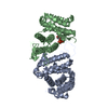
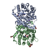
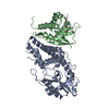
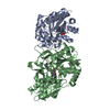
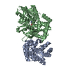
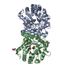
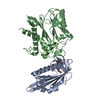
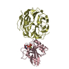
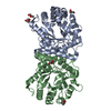
| Title | The crystal structure of native Trypanosoma cruzi dUTPase | ||||||
 Components Components | DEOXYURIDINE TRIPHOSPHATASE DUTP diphosphatase DUTP diphosphatase | ||||||
 Keywords Keywords | HYDROLASE / DUTPASE / TRYPANOSOMA CRUZI / NATIVE / DIMER | ||||||
| Function / homology | all-alpha NTP pyrophosphatase / Type II deoxyuridine triphosphatase / all-alpha NTP pyrophosphatases / Type II deoxyuridine triphosphatase / dUTPase/dCTP pyrophosphatase / dUTPase / Up-down Bundle / Mainly Alpha / Deoxyuridine triphosphatase Function and homology information Function and homology information | ||||||
| Biological species |  TRYPANOSOMA CRUZI (eukaryote) TRYPANOSOMA CRUZI (eukaryote) | ||||||
| Method | X-RAY DIFFRACTION / SYNCHROTRON / MAD / Resolution: 2.4 Å | ||||||
 Authors Authors | Harkiolaki, M. / Dodson, E.J. / Bernier-Villamor, V. / Turkenburg, J.P. / Gonzalez-Pacanowska, D. / Wilson, K.S. | ||||||
 Citation Citation | Journal: Structure / Year: 2004 Title: The Crystal Structure of Trypanosoma Cruzi Dutpase Reveals a Novel Dutp/Dudp Binding Fold Authors: Harkiolaki, M. / Dodson, E.J. / Bernier-Villamor, V. / Turkenburg, J.P. / Gonzalez-Pacanowska, D. / Wilson, K.S. | ||||||
| History |
|
- Structure visualization
| Structure viewer | Molecule: MolmilJmol/JSmol |
|---|
- Downloads & links
-Download
| PDBx/mmCIF format | 1ogl.cif.gz | 57.4 KB | Display | PDBx/mmCIF format |
|---|---|---|---|---|
| PDB format | pdb1ogl.ent.gz | 45.2 KB | Display | PDB format |
| PDBx/mmJSON format | 1ogl.json.gz | Tree view | PDBx/mmJSON format | |
| Others |  Other downloads Other downloads |
-Validation report
| Arichive directory | https://data.pdbj.org/pub/pdb/validation_reports/og/1oglftp://data.pdbj.org/pub/pdb/validation_reports/og/1ogl | HTTPS FTP |
|---|
-Related structure data


-Links
 PDBj
PDBj- Assembly
| Deposited unit | 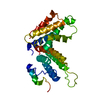
| ||||||||
|---|---|---|---|---|---|---|---|---|---|
| 1 | 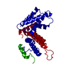
| ||||||||
| Unit cell |
|
-Components
| #1: Protein | DUTP diphosphatase / DUTPASE Mass: 32104.342 Da / Num. of mol.: 1 Source method: isolated from a genetically manipulated source Source: (gene. exp.) TRYPANOSOMA CRUZI (eukaryote) / Strain: Y / Plasmid: PET-11C / Production host:  ESCHERICHIA COLI (E. coli) / Strain (production host): BL21(DE3) / References: UniProt: O15923, dUTP diphosphatase ESCHERICHIA COLI (E. coli) / Strain (production host): BL21(DE3) / References: UniProt: O15923, dUTP diphosphatase |
|---|---|
| #2: Water | ChemComp-HOH / Water Mass: 18.015 Da / Num. of mol.: 34 / Source method: isolated from a natural source / Formula: H2O Mass: 18.015 Da / Num. of mol.: 34 / Source method: isolated from a natural source / Formula: H2O |
-Experimental details
-Experiment
| Experiment | Method: X-RAY DIFFRACTION / Number of used crystals: 1 |
|---|
- Sample preparation
| Crystal | Density Matthews: 2.9 Å3/Da / Density % sol: 57 % Description: ADDITIONAL PHASE INFORMATION PROVIDED THROUGH MERCURYL DERIVATIVE | |||||||||||||||||||||||||||||||||||
|---|---|---|---|---|---|---|---|---|---|---|---|---|---|---|---|---|---|---|---|---|---|---|---|---|---|---|---|---|---|---|---|---|---|---|---|---|
| Crystal grow | pH: 6.6 Details: 15% PEG 2000 MME, 0.1 M LISO4, 50 MM SODIUM CACODYLATE PH 6.6 | |||||||||||||||||||||||||||||||||||
| Crystal grow | *PLUS Temperature: 290 K / pH: 6.6 / Method: vapor diffusion, hanging drop / Details: Harkiolaki, M., (2001) Acta Cryst., D57, 915. | |||||||||||||||||||||||||||||||||||
| Components of the solutions | *PLUS
|
-Data collection
| Diffraction | Mean temperature: 100 K |
|---|---|
| Diffraction source | Source: SYNCHROTRON / Site: ESRF  / Beamline: ID14-4 / Wavelength: 0.9688 / Beamline: ID14-4 / Wavelength: 0.9688 |
| Detector | Type: ADSC CCD / Detector: CCD / Date: Mar 15, 2000 / Details: MIRRORS |
| Radiation | Monochromator: SI111 / Protocol: SINGLE WAVELENGTH / Monochromatic (M) / Laue (L): M / Scattering type: x-ray |
| Radiation wavelength | Wavelength: 0.9688 Å / Relative weight: 1 |
| Reflection | Resolution: 2.4→12 Å / Num. obs: 14827 / % possible obs: 97.5 % / Observed criterion σ(I): 0 / Redundancy: 7.2 % / Rmerge(I) obs: 0.05 / Net I/σ(I): 32.4 |
| Reflection shell | Resolution: 2.4→2.44 Å / Redundancy: 6.5 % / Rmerge(I) obs: 0.41 / Mean I/σ(I) obs: 4.1 / % possible all: 88.4 |
| Reflection | *PLUS Highest resolution: 2.4 Å / Lowest resolution: 12 Å / Redundancy: 7.2 % / Rmerge(I) obs: 0.051 |
| Reflection shell | *PLUS % possible obs: 88.4 % / Rmerge(I) obs: 0.411 / Mean I/σ(I) obs: 4.1 |
- Processing
| Software |
| ||||||||||||||||||||||||||||||||||||||||||||||||||||||||||||||||||||||||||||||||||||||||||||||||||||||||||||||||||||||||||||||||||||||||||||||||||||||||||||||||||||||||||||||||||||||
|---|---|---|---|---|---|---|---|---|---|---|---|---|---|---|---|---|---|---|---|---|---|---|---|---|---|---|---|---|---|---|---|---|---|---|---|---|---|---|---|---|---|---|---|---|---|---|---|---|---|---|---|---|---|---|---|---|---|---|---|---|---|---|---|---|---|---|---|---|---|---|---|---|---|---|---|---|---|---|---|---|---|---|---|---|---|---|---|---|---|---|---|---|---|---|---|---|---|---|---|---|---|---|---|---|---|---|---|---|---|---|---|---|---|---|---|---|---|---|---|---|---|---|---|---|---|---|---|---|---|---|---|---|---|---|---|---|---|---|---|---|---|---|---|---|---|---|---|---|---|---|---|---|---|---|---|---|---|---|---|---|---|---|---|---|---|---|---|---|---|---|---|---|---|---|---|---|---|---|---|---|---|---|---|
| Refinement | Method to determine structure: MAD Starting model: LOW RESOLUTION MAD STRUCTURE Resolution: 2.4→30 Å / Cor.coef. Fo:Fc: 0.951 / Cor.coef. Fo:Fc free: 0.925 / SU B: 7.775 / SU ML: 0.179 / Cross valid method: THROUGHOUT / ESU R: 0.292 / ESU R Free: 0.246 / Stereochemistry target values: MAXIMUM LIKELIHOOD / Details: HYDROGENS HAVE BEEN ADDED IN THE RIDING POSITIONS
| ||||||||||||||||||||||||||||||||||||||||||||||||||||||||||||||||||||||||||||||||||||||||||||||||||||||||||||||||||||||||||||||||||||||||||||||||||||||||||||||||||||||||||||||||||||||
| Solvent computation | Ion probe radii: 0.8 Å / Shrinkage radii: 0.8 Å / VDW probe radii: 1.4 Å / Solvent model: BABINET MODEL WITH MASK | ||||||||||||||||||||||||||||||||||||||||||||||||||||||||||||||||||||||||||||||||||||||||||||||||||||||||||||||||||||||||||||||||||||||||||||||||||||||||||||||||||||||||||||||||||||||
| Displacement parameters | Biso mean: 29.7 Å2
| ||||||||||||||||||||||||||||||||||||||||||||||||||||||||||||||||||||||||||||||||||||||||||||||||||||||||||||||||||||||||||||||||||||||||||||||||||||||||||||||||||||||||||||||||||||||
| Refinement step | Cycle: LAST / Resolution: 2.4→30 Å
| ||||||||||||||||||||||||||||||||||||||||||||||||||||||||||||||||||||||||||||||||||||||||||||||||||||||||||||||||||||||||||||||||||||||||||||||||||||||||||||||||||||||||||||||||||||||
| Refine LS restraints |
|