 Movie
Movie Controller

[English] 日本語
 Yorodumi
- PDB-1oe0: CRYSTAL STRUCTURE OF DROSOPHILA DEOXYRIBONUCLEOSIDE KINASE IN COM... -
+ Open data

- Basic information
| Entry | Database: PDB / ID: 1oe0 | ||||||
|---|---|---|---|---|---|---|---|
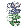
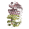
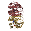
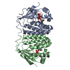
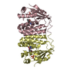
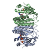
| Title | CRYSTAL STRUCTURE OF DROSOPHILA DEOXYRIBONUCLEOSIDE KINASE IN COMPLEX WITH DTTP | ||||||
 Components Components | DEOXYRIBONUCLEOSIDE KINASE | ||||||
 Keywords Keywords |  TRANSFERASE / DROSOPHILA / DEOXYRIBONUCLEOSIDE KINASE / DTTP / COMPLEX / FEEDBACK INHIBITION / SALVAGE PATHWAY TRANSFERASE / DROSOPHILA / DEOXYRIBONUCLEOSIDE KINASE / DTTP / COMPLEX / FEEDBACK INHIBITION / SALVAGE PATHWAY | ||||||
| Function / homology |  Function and homology informationdeoxynucleoside kinase / Pyrimidine salvage / deoxynucleoside kinase activity / nucleoside salvage / uridine kinase activity / deoxycytidine kinase activity / nucleoside phosphate biosynthetic process / thymidine kinase activity / deoxyguanosine kinase activity / deoxyadenosine kinase activity ...deoxynucleoside kinase / Pyrimidine salvage / deoxynucleoside kinase activity / nucleoside salvage / uridine kinase activity / deoxycytidine kinase activity / nucleoside phosphate biosynthetic process / thymidine kinase activity / deoxyguanosine kinase activity / deoxyadenosine kinase activity / cytidine kinase activity / DNA biosynthetic process / kinase activity / phosphorylation / mitochondrion / ATP binding / cytoplasm Function and homology informationdeoxynucleoside kinase / Pyrimidine salvage / deoxynucleoside kinase activity / nucleoside salvage / uridine kinase activity / deoxycytidine kinase activity / nucleoside phosphate biosynthetic process / thymidine kinase activity / deoxyguanosine kinase activity / deoxyadenosine kinase activity ...deoxynucleoside kinase / Pyrimidine salvage / deoxynucleoside kinase activity / nucleoside salvage / uridine kinase activity / deoxycytidine kinase activity / nucleoside phosphate biosynthetic process / thymidine kinase activity / deoxyguanosine kinase activity / deoxyadenosine kinase activity / cytidine kinase activity / DNA biosynthetic process / kinase activity / phosphorylation / mitochondrion / ATP binding / cytoplasmSimilarity search - Function | ||||||
| Biological species |  DROSOPHILA MELANOGASTER (fruit fly) DROSOPHILA MELANOGASTER (fruit fly) | ||||||
| Method | X-RAY DIFFRACTION / SYNCHROTRON / MOLECULAR REPLACEMENT / Resolution: 2.4 Å | ||||||
 Authors Authors | Mikkelsen, N.E. / Johansson, K. / Karlsson, A. / Knecht, W. / Andersen, G. / Piskur, J. / Munch-Petersen, B. / Eklund, H. | ||||||
 Citation Citation | Journal: Biochemistry / Year: 2003 Title: Structural Basis for Feedback Inhibition of the Deoxyribonucleoside Salvage Pathway:Studies of the Drosophila Deoxyribonucleoside Kinase Authors: Mikkelsen, N.E. / Johansson, K. / Karlsson, A. / Knecht, W. / Andersen, G. / Piskur, J. / Munch-Petersen, B. / Eklund, H. | ||||||
| History |
|
- Structure visualization
| Structure viewer | Molecule: MolmilJmol/JSmol |
|---|
- Downloads & links
-Download
| PDBx/mmCIF format | 1oe0.cif.gz | 182.1 KB | Display | PDBx/mmCIF format |
|---|---|---|---|---|
| PDB format | pdb1oe0.ent.gz | 144.2 KB | Display | PDB format |
| PDBx/mmJSON format | 1oe0.json.gz | Tree view | PDBx/mmJSON format | |
| Others |  Other downloads Other downloads |
-Validation report
| Arichive directory | https://data.pdbj.org/pub/pdb/validation_reports/oe/1oe0ftp://data.pdbj.org/pub/pdb/validation_reports/oe/1oe0 | HTTPS FTP |
|---|
-Related structure data
| Related structure data | 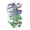 1ot3C 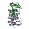 1j90S S: Starting model for refinement C: citing same article ( |
|---|---|
| Similar structure data |

-Links
 PDBj
PDBj
- Assembly
| Deposited unit | 
| ||||||||||||||||
|---|---|---|---|---|---|---|---|---|---|---|---|---|---|---|---|---|---|
| 1 | 
| ||||||||||||||||
| 2 | 
| ||||||||||||||||
| Unit cell |
| ||||||||||||||||
| Noncrystallographic symmetry (NCS) | NCS oper:
|
-Components
| #1: Protein | Mass: 26906.707 Da / Num. of mol.: 4 / Fragment: TRUNCATION MUTANT, RESIDUES 1-230 Source method: isolated from a genetically manipulated source Source: (gene. exp.) DROSOPHILA MELANOGASTER (fruit fly) / Production host:  ESCHERICHIA COLI (E. coli) / Strain (production host): BL21 / References: UniProt: Q9XZT6, deoxynucleoside kinase ESCHERICHIA COLI (E. coli) / Strain (production host): BL21 / References: UniProt: Q9XZT6, deoxynucleoside kinase#2: Chemical | ChemComp-TTP / Thymidine triphosphate  Mass: 482.168 Da / Num. of mol.: 4 / Source method: obtained synthetically / Formula: C10H17N2O14P3 Mass: 482.168 Da / Num. of mol.: 4 / Source method: obtained synthetically / Formula: C10H17N2O14P3#3: Chemical | ChemComp-MG /   Mass: 24.305 Da / Num. of mol.: 4 / Source method: obtained synthetically / Formula: Mg Mass: 24.305 Da / Num. of mol.: 4 / Source method: obtained synthetically / Formula: Mg#4: Water | ChemComp-HOH / | Water Mass: 18.015 Da / Num. of mol.: 169 / Source method: isolated from a natural source / Formula: H2O Mass: 18.015 Da / Num. of mol.: 169 / Source method: isolated from a natural source / Formula: H2O |
|---|
-Experimental details
-Experiment
| Experiment | Method: X-RAY DIFFRACTION / Number of used crystals: 1 |
|---|
- Sample preparation
| Crystal | Density Matthews: 2.78 Å3/Da / Density % sol: 55.8 % |
|---|---|
| Crystal grow | pH: 6.5 / Details: pH 6.50 |
-Data collection
| Diffraction | Mean temperature: 100 K |
|---|---|
| Diffraction source | Source: SYNCHROTRON / Site: ESRF  / Beamline: ID14-2 / Wavelength: 0.933 / Beamline: ID14-2 / Wavelength: 0.933 |
| Detector | Type: MARRESEARCH / Detector: CCD / Date: Apr 18, 2001 |
| Radiation | Protocol: SINGLE WAVELENGTH / Monochromatic (M) / Laue (L): M / Scattering type: x-ray |
| Radiation wavelength | Wavelength: 0.933 Å / Relative weight: 1 |
| Reflection | Resolution: 2.4→25 Å / Num. obs: 42341 / % possible obs: 99.9 % / Redundancy: 5.5 % / Biso Wilson estimate: 37 Å2 / Rmerge(I) obs: 0.062 / Net I/σ(I): 22 |
| Reflection shell | Resolution: 2.4→2.49 Å / Redundancy: 3 % / Rmerge(I) obs: 0.23 / Mean I/σ(I) obs: 4.2 / % possible all: 98.9 |
- Processing
| Software |
| ||||||||||||||||||||||||||||||||||||||||||||||||||||||||||||
|---|---|---|---|---|---|---|---|---|---|---|---|---|---|---|---|---|---|---|---|---|---|---|---|---|---|---|---|---|---|---|---|---|---|---|---|---|---|---|---|---|---|---|---|---|---|---|---|---|---|---|---|---|---|---|---|---|---|---|---|---|---|
| Refinement | Method to determine structure: MOLECULAR REPLACEMENT Starting model: PDB ENTRY 1J90 Resolution: 2.4→24.73 Å / Rfactor Rfree error: 0.005 / Isotropic thermal model: RESTRAINED / Cross valid method: THROUGHOUT / σ(F): 0
| ||||||||||||||||||||||||||||||||||||||||||||||||||||||||||||
| Solvent computation | Solvent model: FLAT MODEL / Bsol: 36.6 Å2 / ksol: 0.33 e/Å3 | ||||||||||||||||||||||||||||||||||||||||||||||||||||||||||||
| Displacement parameters | Biso mean: 55.1 Å2
| ||||||||||||||||||||||||||||||||||||||||||||||||||||||||||||
| Refine analyze |
| ||||||||||||||||||||||||||||||||||||||||||||||||||||||||||||
| Refinement step | Cycle: LAST / Resolution: 2.4→24.73 Å
| ||||||||||||||||||||||||||||||||||||||||||||||||||||||||||||
| Refine LS restraints |
| ||||||||||||||||||||||||||||||||||||||||||||||||||||||||||||
| LS refinement shell | Resolution: 2.4→2.55 Å / Rfactor Rfree error: 0.016 / Total num. of bins used: 6
| ||||||||||||||||||||||||||||||||||||||||||||||||||||||||||||
| Xplor file |
|
