 Movie
Movie Controller

[English] 日本語
 Yorodumi
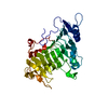
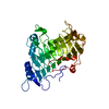
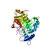
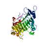
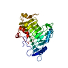
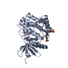
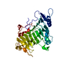
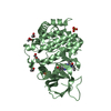
- PDB-1o8e: Pectate Lyase C from Erwinia Chrysanthemi at pH 11.2 with 1mM Ca2+ -
+ Open data

- Basic information
| Entry | Database: PDB / ID: 1o8e | ||||||
|---|---|---|---|---|---|---|---|
| Title | Pectate Lyase C from Erwinia Chrysanthemi at pH 11.2 with 1mM Ca2+ | ||||||
 Components Components | PECTATE LYASE C | ||||||
 Keywords Keywords |  HYDROLASE / PECTATE LYASE CLEAVAGE / CALCIUM BINDING / PARALLEL BETA-HELIX HYDROLASE / PECTATE LYASE CLEAVAGE / CALCIUM BINDING / PARALLEL BETA-HELIX | ||||||
| Function / homology |  Function and homology informationpectate lyase / pectate lyase activity / pectin catabolic process / extracellular region / metal ion binding Function and homology informationpectate lyase / pectate lyase activity / pectin catabolic process / extracellular region / metal ion bindingSimilarity search - Function | ||||||
| Biological species |  ERWINIA CHRYSANTHEMI (bacteria) ERWINIA CHRYSANTHEMI (bacteria) | ||||||
| Method | X-RAY DIFFRACTION / MOLECULAR REPLACEMENT / Resolution: 2.2 Å | ||||||
 Authors Authors | Herron, S.R. / Jurnak, F.A. | ||||||
 Citation Citation | Journal: J.Biol.Chem. / Year: 2003 Title: Characterization and Implications of Ca2+ Binding to Pectate Lyase C Authors: Herron, S.R. / Scavetta, R. / Garrett, M. / Legner, M. / Jurnak, F.A. | ||||||
| History |
|
- Structure visualization
| Structure viewer | Molecule: MolmilJmol/JSmol |
|---|
- Downloads & links
-Download
| PDBx/mmCIF format | 1o8e.cif.gz | 84 KB | Display | PDBx/mmCIF format |
|---|---|---|---|---|
| PDB format | pdb1o8e.ent.gz | 61.5 KB | Display | PDB format |
| PDBx/mmJSON format | 1o8e.json.gz | Tree view | PDBx/mmJSON format | |
| Others |  Other downloads Other downloads |
-Validation report
| Arichive directory | https://data.pdbj.org/pub/pdb/validation_reports/o8/1o8eftp://data.pdbj.org/pub/pdb/validation_reports/o8/1o8e | HTTPS FTP |
|---|
-Related structure data
| Related structure data |  1o88C  1o8dC  1o8fC  1o8gC  1o8hC  1o8iC  1o8jC  1o8kC  1o8lC  1o8mC  1airS S: Starting model for refinement C: citing same article ( |
|---|---|
| Similar structure data |


-Links
 PDBj
PDBj- Assembly
| Deposited unit | 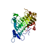
| ||||||||
|---|---|---|---|---|---|---|---|---|---|
| 1 |
| ||||||||
| Unit cell |
|
-Components
| #1: Protein | Mass: 37733.605 Da / Num. of mol.: 1 Source method: isolated from a genetically manipulated source Source: (gene. exp.) ERWINIA CHRYSANTHEMI (bacteria) / Production host: ESCHERICHIA COLI (E. coli) / References: UniProt: P11073, pectate lyase |
|---|---|
| #2: Chemical | ChemComp-CA /   Mass: 40.078 Da / Num. of mol.: 1 / Source method: obtained synthetically / Formula: Ca Mass: 40.078 Da / Num. of mol.: 1 / Source method: obtained synthetically / Formula: Ca |
| #3: Water | ChemComp-HOH / Water Mass: 18.015 Da / Num. of mol.: 182 / Source method: isolated from a natural source / Formula: H2O Mass: 18.015 Da / Num. of mol.: 182 / Source method: isolated from a natural source / Formula: H2O |
-Experimental details
-Experiment
| Experiment | Method: X-RAY DIFFRACTION / Number of used crystals: 1 |
|---|
- Sample preparation
| Crystal | Density Matthews: 3.34 Å3/Da / Density % sol: 63.17 % | ||||||||||||||||||||||||||||||||||||||||||
|---|---|---|---|---|---|---|---|---|---|---|---|---|---|---|---|---|---|---|---|---|---|---|---|---|---|---|---|---|---|---|---|---|---|---|---|---|---|---|---|---|---|---|---|
| Crystal grow | pH: 11.2 / Details: pH 11.20 | ||||||||||||||||||||||||||||||||||||||||||
| Crystal grow | *PLUS Temperature: 4 ℃ / pH: 6.9 / Method: vapor diffusion, sitting drop / Details: Yoder, M. D., (1990) J. Biol. Chem., 265, 11429. | ||||||||||||||||||||||||||||||||||||||||||
| Components of the solutions | *PLUS
|
-Data collection
| Diffraction | Mean temperature: 293 K |
|---|---|
| Diffraction source | Source: ROTATING ANODE / Type: RIGAKU RT3000 / Wavelength: 1.54 |
| Detector | Type: SDMS / Detector: AREA DETECTOR / Details: COLLIMATOR |
| Radiation | Monochromator: GRAPHITE CRYSTAL / Protocol: SINGLE WAVELENGTH / Monochromatic (M) / Laue (L): M / Scattering type: x-ray |
| Radiation wavelength | Wavelength: 1.54 Å / Relative weight: 1 |
| Reflection | Resolution: 2.2→10 Å / Num. obs: 27422 / % possible obs: 92.3 % / Observed criterion σ(I): 2 / Redundancy: 4.1 % / Rmerge(I) obs: 0.076 / Net I/σ(I): 9.6 |
| Reflection | *PLUS Num. measured all: 99422 / Rmerge(I) obs: 0.076 |
- Processing
| Software |
| ||||||||||||||||||||||||||||||||||||||||||||||||||||||||||||
|---|---|---|---|---|---|---|---|---|---|---|---|---|---|---|---|---|---|---|---|---|---|---|---|---|---|---|---|---|---|---|---|---|---|---|---|---|---|---|---|---|---|---|---|---|---|---|---|---|---|---|---|---|---|---|---|---|---|---|---|---|---|
| Refinement | Method to determine structure: MOLECULAR REPLACEMENT Starting model: PDB ENTRY 1AIR Resolution: 2.2→10 Å / Data cutoff high absF: 10000000 / Data cutoff low absF: 0.001 / Cross valid method: THROUGHOUT / σ(F): 2
| ||||||||||||||||||||||||||||||||||||||||||||||||||||||||||||
| Refinement step | Cycle: LAST / Resolution: 2.2→10 Å
| ||||||||||||||||||||||||||||||||||||||||||||||||||||||||||||
| Refine LS restraints |
| ||||||||||||||||||||||||||||||||||||||||||||||||||||||||||||
| Refinement | *PLUS % reflection Rfree: 10 % / Rfactor Rfree: 0.242 | ||||||||||||||||||||||||||||||||||||||||||||||||||||||||||||
| Solvent computation | *PLUS | ||||||||||||||||||||||||||||||||||||||||||||||||||||||||||||
| Displacement parameters | *PLUS | ||||||||||||||||||||||||||||||||||||||||||||||||||||||||||||
| Refine LS restraints | *PLUS
|
