 Movie
Movie Controller


+ Open data

- Basic information
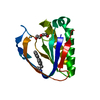
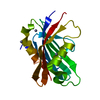
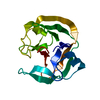
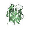
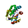
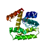
| Entry | Database: PDB / ID: 1o7z | ||||||
|---|---|---|---|---|---|---|---|
| Title | Crystal structure of IP-10 T-form | ||||||
 Components Components | SMALL INDUCIBLE CYTOKINE B10 | ||||||
 Keywords Keywords |  CHEMOKINE / INTERFERON INDUCTION / CHEMOTAXIS / INFLAMMATORY RESPONSE CHEMOKINE / INTERFERON INDUCTION / CHEMOTAXIS / INFLAMMATORY RESPONSE | ||||||
| Function / homology |  Function and homology information Function and homology informationregulation of T cell chemotaxis / negative regulation of myoblast fusion / CXCR3 chemokine receptor binding / regulation of endothelial tube morphogenesis / cellular response to interleukin-17 / response to auditory stimulus / CXCR chemokine receptor binding / cAMP-dependent protein kinase regulator activity / response to vitamin D / negative regulation of myoblast differentiation ...regulation of T cell chemotaxis / negative regulation of myoblast fusion / CXCR3 chemokine receptor binding / regulation of endothelial tube morphogenesis / cellular response to interleukin-17 / response to auditory stimulus / CXCR chemokine receptor binding / cAMP-dependent protein kinase regulator activity / response to vitamin D / negative regulation of myoblast differentiation / T cell chemotaxis / chemokine-mediated signaling pathway / blood circulation / positive regulation of monocyte chemotaxis / Chemokine receptors bind chemokines / endothelial cell activation / chemokine activity / muscle organ development / chemoattractant activity / Interleukin-10 signaling / antiviral innate immune response / positive regulation of T cell migration / negative regulation of angiogenesis / neutrophil chemotaxis / positive regulation of release of sequestered calcium ion into cytosol / response to gamma radiation / adenylate cyclase-activating G protein-coupled receptor signaling pathway / cellular response to virus / chemotaxis / antimicrobial humoral immune response mediated by antimicrobial peptide / cell-cell signaling / cellular response to heat / heparin binding / regulation of cell population proliferation / G alpha (i) signalling events / regulation of apoptotic process / cellular response to lipopolysaccharide / cell surface receptor signaling pathway / inflammatory response / G protein-coupled receptor signaling pathway / external side of plasma membrane / signaling receptor binding / positive regulation of cell population proliferation / signal transduction / positive regulation of transcription by RNA polymerase II / extracellular space / extracellular regionSimilarity search - Function | ||||||
| Biological species |  HOMO SAPIENS (human) HOMO SAPIENS (human) | ||||||
| Method | X-RAY DIFFRACTION / SYNCHROTRON / MOLECULAR REPLACEMENT / Resolution: 1.92 Å | ||||||
 Authors Authors | Swaminathan, G.J. / Holloway, D.E. / Papageorgiou, A.C. / Acharya, K.R. | ||||||
 Citation Citation | Journal: Structure / Year: 2003 Title: Crystal Structures of Oligomeric Forms of the Ip-10/Cxcl10 Chemokine Authors: Swaminathan, G.J. / Holloway, D.E. / Colvin, R.A. / Campanella, G.K. / Papageorgiou, A.C. / Luster, A.D. / Acharya, K.R. | ||||||
| History |
|
- Structure visualization
| Structure viewer | Molecule: MolmilJmol/JSmol |
|---|
- Downloads & links
-Download
| PDBx/mmCIF format | 1o7z.cif.gz | 34.6 KB | Display | PDBx/mmCIF format |
|---|---|---|---|---|
| PDB format | pdb1o7z.ent.gz | 23.9 KB | Display | PDB format |
| PDBx/mmJSON format | 1o7z.json.gz | Tree view | PDBx/mmJSON format | |
| Others |  Other downloads Other downloads |
-Validation report
| Arichive directory | https://data.pdbj.org/pub/pdb/validation_reports/o7/1o7zftp://data.pdbj.org/pub/pdb/validation_reports/o7/1o7z | HTTPS FTP |
|---|
-Related structure data
| Related structure data |  1o7yC 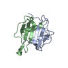 1o80C 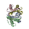 1rhpS C: citing same article ( S: Starting model for refinement |
|---|---|
| Similar structure data |

-Links
 PDBj
PDBj

- Assembly
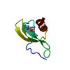
| Deposited unit | 
| ||||||||
|---|---|---|---|---|---|---|---|---|---|
| 1 |
| ||||||||
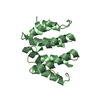
| Unit cell |
|
-Components
| #1: Protein | Mass: 8637.345 Da / Num. of mol.: 2 / Source method: obtained synthetically / Source: (synth.) HOMO SAPIENS (human) / References: UniProt: P02778#2: Water | ChemComp-HOH / | Water Mass: 18.015 Da / Num. of mol.: 42 / Source method: isolated from a natural source / Formula: H2O Mass: 18.015 Da / Num. of mol.: 42 / Source method: isolated from a natural source / Formula: H2OCompound details | CHEMOTACTIC FOR MONOCYTES AND T LYMPHOCYTES. BINDS TO CXCR3. INDUCED BY INTERFERON GAMMA. A DIVERSE ...CHEMOTACTI | Sequence details | THE SEQUENCE CONFLICT INDICATED IN THE SEQADV RECORDS ARISES FROM A DIFFERENCE IN THE PRIMARY ...THE SEQUENCE CONFLICT INDICATED IN THE SEQADV RECORDS ARISES FROM A DIFFERENCE | |
|---|
-Experimental details
-Experiment
| Experiment | Method: X-RAY DIFFRACTION / Number of used crystals: 1 |
|---|
- Sample preparation
| Crystal | Density Matthews: 3.12 Å3/Da / Density % sol: 60.3 % | ||||||||||||||||||||||||
|---|---|---|---|---|---|---|---|---|---|---|---|---|---|---|---|---|---|---|---|---|---|---|---|---|---|
| Crystal grow | pH: 8.75 Details: 10MG/ML PROTEIN,0.1M TRIS-HCL BUFFER, PH 8.75,3.3M SODIUM FORMATE | ||||||||||||||||||||||||
| Crystal grow | *PLUS Temperature: 16 ℃ / Method: vapor diffusion, hanging drop | ||||||||||||||||||||||||
| Components of the solutions | *PLUS
|
-Data collection
| Diffraction | Mean temperature: 100 K |
|---|---|
| Diffraction source | Source: SYNCHROTRON / Site: SRS  / Beamline: PX14.1 / Wavelength: 1.488 / Beamline: PX14.1 / Wavelength: 1.488 |
| Detector | Type: ADSC CCD / Detector: CCD / Date: Feb 25, 2002 / Details: MIRRORS |
| Radiation | Monochromator: SI(111) / Protocol: SINGLE WAVELENGTH / Monochromatic (M) / Laue (L): M / Scattering type: x-ray |
| Radiation wavelength | Wavelength: 1.488 Å / Relative weight: 1 |
| Reflection | Resolution: 1.92→50 Å / Num. obs: 17435 / % possible obs: 99.8 % / Redundancy: 17.8 % / Biso Wilson estimate: 46.8 Å2 / Rmerge(I) obs: 0.087 / Net I/σ(I): 21.7 |
| Reflection shell | Resolution: 1.92→1.97 Å / Rmerge(I) obs: 0.474 / Mean I/σ(I) obs: 3.2 / % possible all: 100 |
| Reflection | *PLUS Lowest resolution: 50 Å / Num. measured all: 309571 |
| Reflection shell | *PLUS Lowest resolution: 1.99 Å / % possible obs: 100 % |
- Processing
| Software |
| ||||||||||||||||||||||||||||||||||||||||||||||||||||||||||||||||||||||||||||||||
|---|---|---|---|---|---|---|---|---|---|---|---|---|---|---|---|---|---|---|---|---|---|---|---|---|---|---|---|---|---|---|---|---|---|---|---|---|---|---|---|---|---|---|---|---|---|---|---|---|---|---|---|---|---|---|---|---|---|---|---|---|---|---|---|---|---|---|---|---|---|---|---|---|---|---|---|---|---|---|---|---|---|
| Refinement | Method to determine structure: MOLECULAR REPLACEMENT Starting model: PDB ENTRY 1RHP Resolution: 1.92→34.58 Å / Rfactor Rfree error: 0.008 / Data cutoff high absF: 874079.2 / Isotropic thermal model: RESTRAINED / Cross valid method: THROUGHOUT / σ(F): 0
| ||||||||||||||||||||||||||||||||||||||||||||||||||||||||||||||||||||||||||||||||
| Solvent computation | Solvent model: FLAT MODEL / Bsol: 52.5121 Å2 / ksol: 0.35472 e/Å3 | ||||||||||||||||||||||||||||||||||||||||||||||||||||||||||||||||||||||||||||||||
| Displacement parameters | Biso mean: 60.1 Å2
| ||||||||||||||||||||||||||||||||||||||||||||||||||||||||||||||||||||||||||||||||
| Refine analyze |
| ||||||||||||||||||||||||||||||||||||||||||||||||||||||||||||||||||||||||||||||||
| Refinement step | Cycle: LAST / Resolution: 1.92→34.58 Å
| ||||||||||||||||||||||||||||||||||||||||||||||||||||||||||||||||||||||||||||||||
| Refine LS restraints |
| ||||||||||||||||||||||||||||||||||||||||||||||||||||||||||||||||||||||||||||||||
| LS refinement shell | Resolution: 1.92→1.99 Å / Rfactor Rfree error: 0.046 / Total num. of bins used: 10
| ||||||||||||||||||||||||||||||||||||||||||||||||||||||||||||||||||||||||||||||||
| Xplor file |
| ||||||||||||||||||||||||||||||||||||||||||||||||||||||||||||||||||||||||||||||||
| Refinement | *PLUS Lowest resolution: 35 Å / % reflection Rfree: 6 % | ||||||||||||||||||||||||||||||||||||||||||||||||||||||||||||||||||||||||||||||||
| Solvent computation | *PLUS | ||||||||||||||||||||||||||||||||||||||||||||||||||||||||||||||||||||||||||||||||
| Displacement parameters | *PLUS | ||||||||||||||||||||||||||||||||||||||||||||||||||||||||||||||||||||||||||||||||
| Refine LS restraints | *PLUS
|
