 Movie
Movie Controller

[English] 日本語
 Yorodumi
- PDB-1nuu: CRYSTAL STRUCTURE OF HUMAN CYTOSOLIC NMN/NaMN ADENYLYLTRANSFERASE... -
+ Open data

- Basic information
| Entry | Database: PDB / ID: 1nuu | ||||||
|---|---|---|---|---|---|---|---|
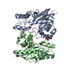
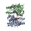
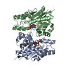
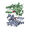
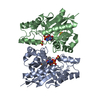
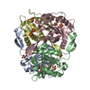
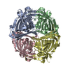
| Title | CRYSTAL STRUCTURE OF HUMAN CYTOSOLIC NMN/NaMN ADENYLYLTRANSFERASE COMPLEXED WITH NAD | ||||||
 Components Components | FKSG76 | ||||||
 Keywords Keywords |  TRANSFERASE / NAD BIOSYNTHESIS / MITOCHONDRIA / PYRIDINE ADENYLYLTRANSFERASE / ENZYME CATALYSIS TRANSFERASE / NAD BIOSYNTHESIS / MITOCHONDRIA / PYRIDINE ADENYLYLTRANSFERASE / ENZYME CATALYSIS | ||||||
| Function / homology |  Function and homology informationnicotinamide-nucleotide adenylyltransferase / nicotinate-nucleotide adenylyltransferase / nicotinamide-nucleotide adenylyltransferase activity / nucleotide biosynthetic process / nicotinate-nucleotide adenylyltransferase activity / Nicotinate metabolism / NAD biosynthetic process / response to tumor necrosis factor / response to wounding / mitochondrial matrix ...nicotinamide-nucleotide adenylyltransferase / nicotinate-nucleotide adenylyltransferase / nicotinamide-nucleotide adenylyltransferase activity / nucleotide biosynthetic process / nicotinate-nucleotide adenylyltransferase activity / Nicotinate metabolism / NAD biosynthetic process / response to tumor necrosis factor / response to wounding / mitochondrial matrix / axon / neuronal cell body / mitochondrion / ATP binding Function and homology informationnicotinamide-nucleotide adenylyltransferase / nicotinate-nucleotide adenylyltransferase / nicotinamide-nucleotide adenylyltransferase activity / nucleotide biosynthetic process / nicotinate-nucleotide adenylyltransferase activity / Nicotinate metabolism / NAD biosynthetic process / response to tumor necrosis factor / response to wounding / mitochondrial matrix ...nicotinamide-nucleotide adenylyltransferase / nicotinate-nucleotide adenylyltransferase / nicotinamide-nucleotide adenylyltransferase activity / nucleotide biosynthetic process / nicotinate-nucleotide adenylyltransferase activity / Nicotinate metabolism / NAD biosynthetic process / response to tumor necrosis factor / response to wounding / mitochondrial matrix / axon / neuronal cell body / mitochondrion / ATP bindingSimilarity search - Function | ||||||
| Biological species |  Homo sapiens (human) Homo sapiens (human) | ||||||
| Method | X-RAY DIFFRACTION / MOLECULAR REPLACEMENT / Resolution: 1.9 Å | ||||||
 Authors Authors | Zhang, X. / Kurnasov, O.V. / Karthikeyan, S. / Grishin, N.V. / Osterman, A.L. / Zhang, H. | ||||||
 Citation Citation | Journal: J.Biol.Chem. / Year: 2003 Title: STRUCTURAL CHARACTERIZATION OF A HUMAN CYTOSOLIC NMN/NaMN ADENYLYLTRANSFERASE AND IMPLICATION IN HUMAN NAD BIOSYNTHESIS Authors: ZHANG, X. / KURNASOV, O.V. / KARTHIKEYAN, S. / GRISHIN, N.V. / OSTERMAN, A.L. / ZHANG, H. #1: Journal: J.Biol.Chem. / Year: 2002Title: STRUCTURE OF HUMAN NICOTINAMIDE/NICOTONIC ACID MONONUCLEOTIDE ADENYLYLTRANSFERASE BASIS FOR THE DUAL SUBSTRATE SPECIFICITY AND ACTIVATION OF THE ONCOLYTIC AGENT TIAZOFURIN Authors: ZHOU, T. / KURNASOV, O. / TOMCHICK, D.R. / BINNS, D.D. / GRISHIN, N.V. / MARQUEZ, V.E. / OSTERMAN, A.L. / ZHANG, H. | ||||||
| History |
|
- Structure visualization
| Structure viewer | Molecule: MolmilJmol/JSmol |
|---|
- Downloads & links
-Download
| PDBx/mmCIF format | 1nuu.cif.gz | 107.2 KB | Display | PDBx/mmCIF format |
|---|---|---|---|---|
| PDB format | pdb1nuu.ent.gz | 81.4 KB | Display | PDB format |
| PDBx/mmJSON format | 1nuu.json.gz | Tree view | PDBx/mmJSON format | |
| Others |  Other downloads Other downloads |
-Validation report
| Arichive directory | https://data.pdbj.org/pub/pdb/validation_reports/nu/1nuuftp://data.pdbj.org/pub/pdb/validation_reports/nu/1nuu | HTTPS FTP |
|---|
-Related structure data
| Related structure data |  1nupC  1nuqC  1nurC  1nusC  1nutC  1kqnS C: citing same article ( S: Starting model for refinement |
|---|---|
| Similar structure data |

-Links
 PDBj
PDBj- Assembly
| Deposited unit | 
| ||||||||
|---|---|---|---|---|---|---|---|---|---|
| 1 | 
| ||||||||
| Unit cell |
| ||||||||
| Details | Biological assembly is tetramer and is generated by two fold axis : Y,X,-Z |
-Components
| #1: Protein | Mass: 28396.768 Da / Num. of mol.: 2 Source method: isolated from a genetically manipulated source Source: (gene. exp.) Homo sapiens (human) / Gene: FKSG76 / Plasmid: pPROEX / Production host:  Escherichia coli (E. coli) / Strain (production host): DH10-beta / References: UniProt: Q96T66 Escherichia coli (E. coli) / Strain (production host): DH10-beta / References: UniProt: Q96T66#2: Chemical | ChemComp-SO4 / Sulfate  Mass: 96.063 Da / Num. of mol.: 4 / Source method: obtained synthetically / Formula: SO4 Mass: 96.063 Da / Num. of mol.: 4 / Source method: obtained synthetically / Formula: SO4#3: Chemical | Nicotinamide adenine dinucleotide  Mass: 663.425 Da / Num. of mol.: 2 / Source method: obtained synthetically / Formula: C21H27N7O14P2 / Comment: NAD*YM Mass: 663.425 Da / Num. of mol.: 2 / Source method: obtained synthetically / Formula: C21H27N7O14P2 / Comment: NAD*YM#4: Water | ChemComp-HOH / | Water Mass: 18.015 Da / Num. of mol.: 268 / Source method: isolated from a natural source / Formula: H2O Mass: 18.015 Da / Num. of mol.: 268 / Source method: isolated from a natural source / Formula: H2O |
|---|
-Experimental details
-Experiment
| Experiment | Method: X-RAY DIFFRACTION / Number of used crystals: 1 |
|---|
- Sample preparation
| Crystal | Density Matthews: 2.18 Å3/Da / Density % sol: 43.26 % | |||||||||||||||||||||||||||||||||||
|---|---|---|---|---|---|---|---|---|---|---|---|---|---|---|---|---|---|---|---|---|---|---|---|---|---|---|---|---|---|---|---|---|---|---|---|---|
| Crystal grow | Temperature: 293 K / Method: vapor diffusion, hanging drop / pH: 6.5 Details: NA CADODALYTE, LITHIUM SULPHATE, PEG 400, pH 6.5, VAPOR DIFFUSION, HANGING DROP, temperature 293K | |||||||||||||||||||||||||||||||||||
| Crystal grow | *PLUS Method: vapor diffusion, hanging drop / PH range low: 7 / PH range high: 6 | |||||||||||||||||||||||||||||||||||
| Components of the solutions | *PLUS
|
-Data collection
| Diffraction | Mean temperature: 100 K |
|---|---|
| Diffraction source | Source: ROTATING ANODE / Type: RIGAKU RUH3R / Wavelength: 1.5418 Å |
| Detector | Type: RIGAKU RAXIS IV / Detector: IMAGE PLATE / Date: Jul 5, 2002 / Details: OSMIC MIRROR |
| Radiation | Monochromator: MIRRORS / Protocol: SINGLE WAVELENGTH / Monochromatic (M) / Laue (L): M / Scattering type: x-ray |
| Radiation wavelength | Wavelength: 1.5418 Å / Relative weight: 1 |
| Reflection | Resolution: 1.9→100 Å / Num. all: 37478 / Num. obs: 36764 / Observed criterion σ(F): 0 / Observed criterion σ(I): 0 / Redundancy: 7.1 % / Biso Wilson estimate: 25.9 Å2 / Rsym value: 0.058 / Net I/σ(I): 34.4 |
| Reflection shell | Resolution: 1.9→1.93 Å / Mean I/σ(I) obs: 3 / Rsym value: 0.468 / % possible all: 91.1 |
| Reflection | *PLUS Num. obs: 37478 / % possible obs: 99 % / Num. measured all: 268063 / Rmerge(I) obs: 0.058 |
| Reflection shell | *PLUS % possible obs: 91.1 % / Rmerge(I) obs: 0.468 |
- Processing
| Software |
| ||||||||||||||||||||||||||||||||||||||||||||||||||||||||||||||||||||||||||||||||
|---|---|---|---|---|---|---|---|---|---|---|---|---|---|---|---|---|---|---|---|---|---|---|---|---|---|---|---|---|---|---|---|---|---|---|---|---|---|---|---|---|---|---|---|---|---|---|---|---|---|---|---|---|---|---|---|---|---|---|---|---|---|---|---|---|---|---|---|---|---|---|---|---|---|---|---|---|---|---|---|---|---|
| Refinement | Method to determine structure: MOLECULAR REPLACEMENT Starting model: PDB ENTRY 1KQN Resolution: 1.9→69.83 Å / Rfactor Rfree error: 0.006 / Data cutoff high absF: 473330.33 / Data cutoff low absF: 0 / Isotropic thermal model: RESTRAINED / Cross valid method: THROUGHOUT / σ(F): 0 / Stereochemistry target values: Engh & Huber
| ||||||||||||||||||||||||||||||||||||||||||||||||||||||||||||||||||||||||||||||||
| Solvent computation | Solvent model: FLAT MODEL / Bsol: 40.8842 Å2 / ksol: 0.353723 e/Å3 | ||||||||||||||||||||||||||||||||||||||||||||||||||||||||||||||||||||||||||||||||
| Displacement parameters | Biso mean: 32.6 Å2
| ||||||||||||||||||||||||||||||||||||||||||||||||||||||||||||||||||||||||||||||||
| Refine analyze |
| ||||||||||||||||||||||||||||||||||||||||||||||||||||||||||||||||||||||||||||||||
| Refinement step | Cycle: LAST / Resolution: 1.9→69.83 Å
| ||||||||||||||||||||||||||||||||||||||||||||||||||||||||||||||||||||||||||||||||
| Refine LS restraints |
| ||||||||||||||||||||||||||||||||||||||||||||||||||||||||||||||||||||||||||||||||
| LS refinement shell | Resolution: 1.9→2.02 Å / Rfactor Rfree error: 0.017 / Total num. of bins used: 6
| ||||||||||||||||||||||||||||||||||||||||||||||||||||||||||||||||||||||||||||||||
| Xplor file |
| ||||||||||||||||||||||||||||||||||||||||||||||||||||||||||||||||||||||||||||||||
| Refinement | *PLUS % reflection Rfree: 5 % / Rfactor Rfree: 0.242 / Rfactor Rwork: 0.198 | ||||||||||||||||||||||||||||||||||||||||||||||||||||||||||||||||||||||||||||||||
| Solvent computation | *PLUS | ||||||||||||||||||||||||||||||||||||||||||||||||||||||||||||||||||||||||||||||||
| Displacement parameters | *PLUS | ||||||||||||||||||||||||||||||||||||||||||||||||||||||||||||||||||||||||||||||||
| Refine LS restraints | *PLUS
|
