 Movie
Movie Controller

[English] 日本語
 Yorodumi
- PDB-1nuk: CRYSTAL STRUCTURE OF THE LIGAND-BINDING DOMAIN OF THE EPHB2 RECEP... -
+ Open data

- Basic information
| Entry | Database: PDB / ID: 1nuk | ||||||
|---|---|---|---|---|---|---|---|
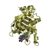
| Title | CRYSTAL STRUCTURE OF THE LIGAND-BINDING DOMAIN OF THE EPHB2 RECEPTOR TYROSINE KINASE | ||||||
 Components Components | PROTEIN (TYROSINE-PROTEIN KINASE RECEPTOR EPH) | ||||||
 Keywords Keywords |  TRANSFERASE / EPH RECEPTOR TYROSINE KINASE TRANSFERASE / EPH RECEPTOR TYROSINE KINASE | ||||||
| Function / homology |  Function and homology information Function and homology informationregulation of T-helper 17 type immune response / trans-synaptic signaling by trans-synaptic complex, modulating synaptic transmission / Ephrin signaling / negative regulation of NMDA glutamate receptor activity / EPH-Ephrin signaling / hindbrain tangential cell migration / vesicle-mediated intercellular transport / EPH-ephrin mediated repulsion of cells / EPHB-mediated forward signaling / positive regulation of NMDA glutamate receptor activity ...regulation of T-helper 17 type immune response / trans-synaptic signaling by trans-synaptic complex, modulating synaptic transmission / Ephrin signaling / negative regulation of NMDA glutamate receptor activity / EPH-Ephrin signaling / hindbrain tangential cell migration / vesicle-mediated intercellular transport / EPH-ephrin mediated repulsion of cells / EPHB-mediated forward signaling / positive regulation of NMDA glutamate receptor activity / postsynaptic membrane assembly / urogenital system development / regulation of body fluid levels / optic nerve morphogenesis / tight junction assembly / neuron projection retraction / axon guidance receptor activity / central nervous system projection neuron axonogenesis / negative regulation of axonogenesis / regulation of behavioral fear response / transmembrane-ephrin receptor activity / positive regulation of long-term neuronal synaptic plasticity / regulation of autophagosome assembly / positive regulation of dendritic spine morphogenesis / dendritic spine development / corpus callosum development / positive regulation of synaptic plasticity / camera-type eye morphogenesis / regulation of filopodium assembly / ephrin receptor activity / negative regulation of Ras protein signal transduction / positive regulation of protein localization to cell surface / commissural neuron axon guidance / dendritic spine morphogenesis / negative regulation of cell adhesion / retinal ganglion cell axon guidance / axonal fasciculation / positive regulation of synapse assembly / regulation of receptor signaling pathway via JAK-STAT / regulation of synapse assembly / inner ear morphogenesis / regulation of neuronal synaptic plasticity / regulation of axonogenesis / B cell activation / roof of mouth development / regulation of blood coagulation / positive regulation of immunoglobulin production / negative regulation of cytokine production involved in inflammatory response / ephrin receptor signaling pathway / positive regulation of B cell proliferation / hippocampal mossy fiber to CA3 synapse / axonogenesis / negative regulation of protein phosphorylation / learning / positive regulation of long-term synaptic potentiation / axon guidance / positive regulation of protein localization to plasma membrane / animal organ morphogenesis / negative regulation of protein kinase activity / cell morphogenesis / negative regulation of ERK1 and ERK2 cascade / receptor protein-tyrosine kinase / peptidyl-tyrosine phosphorylation / cellular response to amyloid-beta / cell surface receptor protein tyrosine kinase signaling pathway / positive regulation of tumor necrosis factor production / signaling receptor activity / presynaptic membrane / amyloid-beta binding / postsynaptic membrane / postsynapse / angiogenesis / protein tyrosine kinase activity / cellular response to lipopolysaccharide / dendritic spine / learning or memory / positive regulation of cell migration / axon / phosphorylation / signaling receptor binding / dendrite / neuronal cell body / glutamatergic synapse / synapse / protein-containing complex binding / positive regulation of gene expression / cell surface / nucleoplasm / ATP binding / identical protein binding / plasma membraneSimilarity search - Function | ||||||
| Biological species |  Mus musculus (house mouse) Mus musculus (house mouse) | ||||||
| Method | X-RAY DIFFRACTION / MIR / Resolution: 2.9 Å | ||||||
 Authors Authors | Himanen, J.-P. / Henkemeyer, M. / Nikolov, D.B. | ||||||
 Citation Citation | Journal: Nature / Year: 1998 Title: Crystal structure of the ligand-binding domain of the receptor tyrosine kinase EphB2. Authors: Himanen, J.P. / Henkemeyer, M. / Nikolov, D.B. | ||||||
| History |
|
- Structure visualization
| Structure viewer | Molecule: MolmilJmol/JSmol |
|---|
- Downloads & links
-Download
| PDBx/mmCIF format | 1nuk.cif.gz | 44.8 KB | Display | PDBx/mmCIF format |
|---|---|---|---|---|
| PDB format | pdb1nuk.ent.gz | 32.4 KB | Display | PDB format |
| PDBx/mmJSON format | 1nuk.json.gz | Tree view | PDBx/mmJSON format | |
| Others |  Other downloads Other downloads |
-Validation report
| Arichive directory | https://data.pdbj.org/pub/pdb/validation_reports/nu/1nukftp://data.pdbj.org/pub/pdb/validation_reports/nu/1nuk | HTTPS FTP |
|---|
-Related structure data
| Similar structure data |
|---|


-Links
 PDBj
PDBj


- Assembly
| Deposited unit | 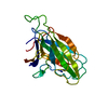
| ||||||||
|---|---|---|---|---|---|---|---|---|---|
| 1 |
| ||||||||
| Unit cell |
|
-Components
| #1: Protein | Mass: 21139.881 Da / Num. of mol.: 1 / Fragment: LIGAND-BINDING DOMAIN Source method: isolated from a genetically manipulated source Source: (gene. exp.) Mus musculus (house mouse) / Production host:  Escherichia coli (E. coli) / Strain (production host): AD494 / References: UniProt: P54763, EC: 2.7.1.112 Escherichia coli (E. coli) / Strain (production host): AD494 / References: UniProt: P54763, EC: 2.7.1.112 |
|---|
-Experimental details
-Experiment
| Experiment | Method: X-RAY DIFFRACTION |
|---|
- Sample preparation
| Crystal | Density Matthews: 2.86 Å3/Da / Density % sol: 56.94 % | ||||||||||||||||||||||||||||||||||||||||||||||||
|---|---|---|---|---|---|---|---|---|---|---|---|---|---|---|---|---|---|---|---|---|---|---|---|---|---|---|---|---|---|---|---|---|---|---|---|---|---|---|---|---|---|---|---|---|---|---|---|---|---|
| Crystal grow | pH: 5 / Details: pH 5.0 | ||||||||||||||||||||||||||||||||||||||||||||||||
| Crystal grow | *PLUS pH: 7.2 / Method: vapor diffusion, hanging drop | ||||||||||||||||||||||||||||||||||||||||||||||||
| Components of the solutions | *PLUS
|
-Data collection
| Diffraction | Mean temperature: 290 K |
|---|---|
| Diffraction source | Source: ROTATING ANODE / Type: RIGAKU RU200 / Wavelength: 1.5418 |
| Radiation | Protocol: SINGLE WAVELENGTH / Monochromatic (M) / Laue (L): M / Scattering type: x-ray |
| Radiation wavelength | Wavelength: 1.5418 Å / Relative weight: 1 |
| Reflection | Resolution: 2.9→8 Å / Num. obs: 4214 / % possible obs: 95 % / Observed criterion σ(I): 2 / Redundancy: 5 % / Rmerge(I) obs: 0.11 / Rsym value: 0.11 |
| Reflection | *PLUS Redundancy: 5 % |
- Processing
| Software | Name: X-PLOR / Version: 3.1 / Classification: refinement | ||||||||||||||||||||||||||||||||||||||||||||||||||||||||||||
|---|---|---|---|---|---|---|---|---|---|---|---|---|---|---|---|---|---|---|---|---|---|---|---|---|---|---|---|---|---|---|---|---|---|---|---|---|---|---|---|---|---|---|---|---|---|---|---|---|---|---|---|---|---|---|---|---|---|---|---|---|---|
| Refinement | Method to determine structure: MIR / Resolution: 2.9→8 Å / σ(F): 2
| ||||||||||||||||||||||||||||||||||||||||||||||||||||||||||||
| Displacement parameters | Biso mean: 24.9 Å2 | ||||||||||||||||||||||||||||||||||||||||||||||||||||||||||||
| Refinement step | Cycle: LAST / Resolution: 2.9→8 Å
| ||||||||||||||||||||||||||||||||||||||||||||||||||||||||||||
| Refine LS restraints |
| ||||||||||||||||||||||||||||||||||||||||||||||||||||||||||||
| Software | *PLUS Version: 3.1 / Classification: refinement | ||||||||||||||||||||||||||||||||||||||||||||||||||||||||||||
| Refinement | *PLUS Highest resolution: 2.9 Å / Lowest resolution: 8 Å / σ(F): 2 / % reflection Rfree: 7.5 % | ||||||||||||||||||||||||||||||||||||||||||||||||||||||||||||
| Solvent computation | *PLUS | ||||||||||||||||||||||||||||||||||||||||||||||||||||||||||||
| Displacement parameters | *PLUS Biso mean: 24.9 Å2 |
