 Movie
Movie Controller

[English] 日本語
 Yorodumi
- PDB-1ntg: Crystal Structure of the EMAP II-like Cytokine Released from huma... -
+ Open data

- Basic information
| Entry | Database: PDB / ID: 1ntg | ||||||
|---|---|---|---|---|---|---|---|
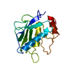
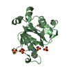
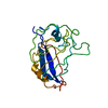
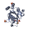
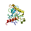
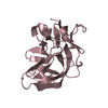
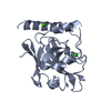
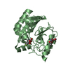
| Title | Crystal Structure of the EMAP II-like Cytokine Released from human tyrosyl-tRNA Synthetase | ||||||
 Components Components | Tyrosyl-tRNA synthetase | ||||||
 Keywords Keywords |  LIGASE / beta barrel LIGASE / beta barrel | ||||||
| Function / homology |  Function and homology informationinterleukin-8 receptor binding / tyrosyl-tRNA aminoacylation / tyrosine-tRNA ligase / tyrosine-tRNA ligase activity / Cytosolic tRNA aminoacylation / response to starvation / small molecule binding / tRNA binding / nuclear body / apoptotic process ...interleukin-8 receptor binding / tyrosyl-tRNA aminoacylation / tyrosine-tRNA ligase / tyrosine-tRNA ligase activity / Cytosolic tRNA aminoacylation / response to starvation / small molecule binding / tRNA binding / nuclear body / apoptotic process / extracellular space / RNA binding / ATP binding / nucleus / cytosol / cytoplasm Function and homology informationinterleukin-8 receptor binding / tyrosyl-tRNA aminoacylation / tyrosine-tRNA ligase / tyrosine-tRNA ligase activity / Cytosolic tRNA aminoacylation / response to starvation / small molecule binding / tRNA binding / nuclear body / apoptotic process ...interleukin-8 receptor binding / tyrosyl-tRNA aminoacylation / tyrosine-tRNA ligase / tyrosine-tRNA ligase activity / Cytosolic tRNA aminoacylation / response to starvation / small molecule binding / tRNA binding / nuclear body / apoptotic process / extracellular space / RNA binding / ATP binding / nucleus / cytosol / cytoplasmSimilarity search - Function | ||||||
| Biological species |  Homo sapiens (human) Homo sapiens (human) | ||||||
| Method | X-RAY DIFFRACTION / MOLECULAR REPLACEMENT / Resolution: 2.21 Å | ||||||
 Authors Authors | Yang, X.-L. / Skene, R.J. / Mcree, D.E. / Schimmel, P. | ||||||
 Citation Citation | Journal: Helv.Chim.Acta / Year: 2003 Title: Crystal Structure of an EMAP-II-like Cytokine Released from a human tRNA Synthetase Authors: Yang, X.-L. / Liu, J. / Skene, R.J. / McRee, D.E. / Schimmel, P. | ||||||
| History |
|
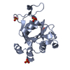
- Structure visualization
| Structure viewer | Molecule: MolmilJmol/JSmol |
|---|
- Downloads & links
-Download
| PDBx/mmCIF format | 1ntg.cif.gz | 142.5 KB | Display | PDBx/mmCIF format |
|---|---|---|---|---|
| PDB format | pdb1ntg.ent.gz | 112.7 KB | Display | PDB format |
| PDBx/mmJSON format | 1ntg.json.gz | Tree view | PDBx/mmJSON format | |
| Others |  Other downloads Other downloads |
-Validation report
| Arichive directory | https://data.pdbj.org/pub/pdb/validation_reports/nt/1ntgftp://data.pdbj.org/pub/pdb/validation_reports/nt/1ntg | HTTPS FTP |
|---|
-Related structure data
| Related structure data |  1fl0S S: Starting model for refinement |
|---|---|
| Similar structure data |
-Links
 PDBj
PDBj
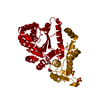
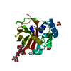
- Assembly
| Deposited unit | 
| ||||||||
|---|---|---|---|---|---|---|---|---|---|
| 1 | 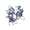
| ||||||||
| 2 | 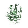
| ||||||||
| 3 | 
| ||||||||
| 4 | 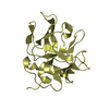
| ||||||||
| Unit cell |
|
-Components
| #1: Protein | Mass: 19124.049 Da / Num. of mol.: 4 / Fragment: C-TyrRS Source method: isolated from a genetically manipulated source Source: (gene. exp.) Homo sapiens (human) / Gene: YARS / Plasmid: PET20b / Production host:  Escherichia coli (E. coli) / References: UniProt: P54577, tyrosine-tRNA ligase Escherichia coli (E. coli) / References: UniProt: P54577, tyrosine-tRNA ligase#2: Water | ChemComp-HOH / | Water Mass: 18.015 Da / Num. of mol.: 182 / Source method: isolated from a natural source / Formula: H2O Mass: 18.015 Da / Num. of mol.: 182 / Source method: isolated from a natural source / Formula: H2O |
|---|
-Experimental details
-Experiment
| Experiment | Method: X-RAY DIFFRACTION / Number of used crystals: 1 |
|---|
- Sample preparation
| Crystal | Density Matthews: 2.63 Å3/Da / Density % sol: 52.85 % | ||||||||||||||||||||||||
|---|---|---|---|---|---|---|---|---|---|---|---|---|---|---|---|---|---|---|---|---|---|---|---|---|---|
| Crystal grow | Temperature: 279 K / Method: vapor diffusion, sitting drop / pH: 6.9 Details: ammonium sulfate, Sodium phosphate mono-basic, potassium phosphate di-basic, acetone, pH 6.9, VAPOR DIFFUSION, SITTING DROP, temperature 279K | ||||||||||||||||||||||||
| Crystal grow | *PLUS Temperature: 4 ℃ | ||||||||||||||||||||||||
| Components of the solutions | *PLUS
|
-Data collection
| Diffraction | Mean temperature: 100 K |
|---|---|
| Diffraction source | Source: ROTATING ANODE / Type: SIEMENS / Wavelength: 1.5418 Å |
| Detector | Type: MARRESEARCH / Detector: AREA DETECTOR / Date: Oct 30, 2001 |
| Radiation | Monochromator: mirror / Protocol: SINGLE WAVELENGTH / Monochromatic (M) / Laue (L): M / Scattering type: x-ray |
| Radiation wavelength | Wavelength: 1.5418 Å / Relative weight: 1 |
| Reflection | Resolution: 2.21→20 Å / Num. all: 39281 / Num. obs: 39281 / % possible obs: 94.6 % / Observed criterion σ(F): 0 / Observed criterion σ(I): 0 / Redundancy: 3.7 % / Rmerge(I) obs: 0.067 |
| Reflection shell | Resolution: 2.21→2.29 Å / Rmerge(I) obs: 0.252 / Mean I/σ(I) obs: 3.8 / % possible all: 83.7 |
| Reflection | *PLUS Highest resolution: 2.2 Å / Lowest resolution: 20 Å |
| Reflection shell | *PLUS % possible obs: 83.7 % |
- Processing
| Software |
| ||||||||||||||||||||
|---|---|---|---|---|---|---|---|---|---|---|---|---|---|---|---|---|---|---|---|---|---|
| Refinement | Method to determine structure: MOLECULAR REPLACEMENT Starting model: EMAP II structure (PDB 1FL0) Resolution: 2.21→20 Å / σ(F): 0 / Stereochemistry target values: ENGH & HUBER
| ||||||||||||||||||||
| Refinement step | Cycle: LAST / Resolution: 2.21→20 Å
| ||||||||||||||||||||
| Refine LS restraints |
| ||||||||||||||||||||
| Software | *PLUS Name: CNS / Classification: refinement | ||||||||||||||||||||
| Refinement | *PLUS Highest resolution: 2.2 Å / Lowest resolution: 20 Å / % reflection Rfree: 5 % | ||||||||||||||||||||
| Solvent computation | *PLUS | ||||||||||||||||||||
| Displacement parameters | *PLUS |
