 Movie
Movie Controller


+ Open data

- Basic information
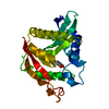
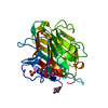
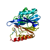
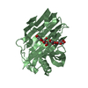
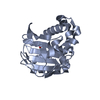
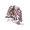
| Entry | Database: PDB / ID: 1nn6 | |||||||||
|---|---|---|---|---|---|---|---|---|---|---|
| Title | Human Pro-Chymase | |||||||||
 Components Components | Chymase | |||||||||
 Keywords Keywords | HYDROLASE / Serine Protease / Zymogen / Chymase / Conformational change | |||||||||
| Function / homology |  Function and homology informationchymase / basement membrane disassembly / cytokine precursor processing / midbrain development / Activation of Matrix Metalloproteinases / angiotensin maturation / extracellular matrix disassembly / Metabolism of Angiotensinogen to Angiotensins / serine-type peptidase activity / secretory granule ...chymase / basement membrane disassembly / cytokine precursor processing / midbrain development / Activation of Matrix Metalloproteinases / angiotensin maturation / extracellular matrix disassembly / Metabolism of Angiotensinogen to Angiotensins / serine-type peptidase activity / secretory granule / cellular response to glucose stimulus / peptide binding / protein catabolic process / Signaling by SCF-KIT / cytoplasmic ribonucleoprotein granule / positive regulation of angiogenesis / regulation of inflammatory response / collagen-containing extracellular matrix / endopeptidase activity / serine-type endopeptidase activity / intracellular membrane-bounded organelle / extracellular space / extracellular region / cytosol / cytoplasm Function and homology informationchymase / basement membrane disassembly / cytokine precursor processing / midbrain development / Activation of Matrix Metalloproteinases / angiotensin maturation / extracellular matrix disassembly / Metabolism of Angiotensinogen to Angiotensins / serine-type peptidase activity / secretory granule ...chymase / basement membrane disassembly / cytokine precursor processing / midbrain development / Activation of Matrix Metalloproteinases / angiotensin maturation / extracellular matrix disassembly / Metabolism of Angiotensinogen to Angiotensins / serine-type peptidase activity / secretory granule / cellular response to glucose stimulus / peptide binding / protein catabolic process / Signaling by SCF-KIT / cytoplasmic ribonucleoprotein granule / positive regulation of angiogenesis / regulation of inflammatory response / collagen-containing extracellular matrix / endopeptidase activity / serine-type endopeptidase activity / intracellular membrane-bounded organelle / extracellular space / extracellular region / cytosol / cytoplasmSimilarity search - Function | |||||||||
| Biological species |  Homo sapiens (human) Homo sapiens (human) | |||||||||
| Method | X-RAY DIFFRACTION / SYNCHROTRON / MOLECULAR REPLACEMENT / Resolution: 1.75 Å | |||||||||
 Authors Authors | Reiling, K.K. / Krucinski, J. / Miercke, L.J.W. / Raymond, W.W. / Caughey, G.H. / Stroud, R.M. | |||||||||
 Citation Citation | Journal: Biochemistry / Year: 2003 Title: Structure of human pro-chymase: a model for the activating transition of granule-associated proteases. Authors: Reiling, K.K. / Krucinski, J. / Miercke, L.J. / Raymond, W.W. / Caughey, G.H. / Stroud, R.M. #1: Journal: Biochemistry / Year: 1997Title: Crystal structure of phenylmethanesulfonyl fluoride-treated human chymase at 1.9 A Authors: McGrath, M.E. / Mirzadegan, T. / Schmidt, B.F. #2: Journal: J.Mol.Biol. / Year: 1999Title: The 2.2 A Crystal Structure of Human Chymase in Complex with Succinyl-Ala-Ala-Pro-Phe-chloromethylketone: Structural Explanation for its Dipeptidyl Carboxypeptidase Specificity Authors: Pereira, P.J. / Wang, Z.M. / Rubin, H. / Huber, R. / Bode, W. / Schechter, N.M. / Strobl, S. #3: Journal: Biochemistry / Year: 1977Title: Structure of bovine trypsinogen at 1.9 A resolution. Authors: Kossiakoff, A.A. / Chambers, J.L. / Kay, L.M. / Stroud, R.M. #4: Journal: J.Biol.Chem. / Year: 1991Title: Structure, chromosomal assignment, and deduced amino acid sequence of a human gene for mast cell chymase. Authors: Caughey, G.H. / Zerweck, E.H. / Vanderslice, P. | |||||||||
| History |
|
- Structure visualization
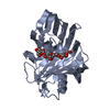
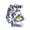
| Structure viewer | Molecule: MolmilJmol/JSmol |
|---|
- Downloads & links
-Download
| PDBx/mmCIF format | 1nn6.cif.gz | 62.9 KB | Display | PDBx/mmCIF format |
|---|---|---|---|---|
| PDB format | pdb1nn6.ent.gz | 44.6 KB | Display | PDB format |
| PDBx/mmJSON format | 1nn6.json.gz | Tree view | PDBx/mmJSON format | |
| Others |  Other downloads Other downloads |
-Validation report
| Arichive directory | https://data.pdbj.org/pub/pdb/validation_reports/nn/1nn6ftp://data.pdbj.org/pub/pdb/validation_reports/nn/1nn6 | HTTPS FTP |
|---|
-Related structure data
| Related structure data | 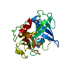 1kltS S: Starting model for refinement |
|---|---|
| Similar structure data |
-Links
 PDBj
PDBj


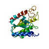
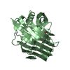
- Assembly
| Deposited unit | 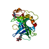
| ||||||||
|---|---|---|---|---|---|---|---|---|---|
| 1 |
| ||||||||
| Unit cell |
|
-Components
| #1: Protein | / Mast cell protease I Mass: 25253.135 Da / Num. of mol.: 1 Source method: isolated from a genetically manipulated source Source: (gene. exp.) Homo sapiens (human) / Gene: Human mast cell chymase gene / Cell line (production host): SF9 / Production host:   Spodoptera frugiperda (fall armyworm) / Strain (production host): High Five (Trichoplusia ni) cells / References: UniProt: P23946, chymase Spodoptera frugiperda (fall armyworm) / Strain (production host): High Five (Trichoplusia ni) cells / References: UniProt: P23946, chymase |
|---|---|
| #2: Polysaccharide | 2-acetamido-2-deoxy-beta-D-glucopyranose-(1-4)-2-acetamido-2-deoxy-beta-D-glucopyranose / Mass: 424.401 Da / Num. of mol.: 1 Source method: isolated from a genetically manipulated source |
| #3: Sugar | ChemComp-NAG / N-Acetylglucosamine  Type: D-saccharide, beta linking / Mass: 221.208 Da / Num. of mol.: 1 Type: D-saccharide, beta linking / Mass: 221.208 Da / Num. of mol.: 1Source method: isolated from a genetically manipulated source Formula: C8H15NO6 |
| #4: Water | ChemComp-HOH / Water Mass: 18.015 Da / Num. of mol.: 153 / Source method: isolated from a natural source / Formula: H2O Mass: 18.015 Da / Num. of mol.: 153 / Source method: isolated from a natural source / Formula: H2O |
-Experimental details
-Experiment
| Experiment | Method: X-RAY DIFFRACTION / Number of used crystals: 1 |
|---|
- Sample preparation
| Crystal | Density Matthews: 2.35 Å3/Da / Density % sol: 47.32 % | |||||||||||||||||||||||||||||||||||||||||||||||||
|---|---|---|---|---|---|---|---|---|---|---|---|---|---|---|---|---|---|---|---|---|---|---|---|---|---|---|---|---|---|---|---|---|---|---|---|---|---|---|---|---|---|---|---|---|---|---|---|---|---|---|
| Crystal grow | Temperature: 277 K / Method: vapor diffusion, hanging drop / pH: 8.5 Details: 20% Peg 4k, 150mM Na Formate, and 100 mM Tris , pH 8.5, VAPOR DIFFUSION, HANGING DROP, temperature 277K | |||||||||||||||||||||||||||||||||||||||||||||||||
| Crystal grow | *PLUS Temperature: 4 ℃ | |||||||||||||||||||||||||||||||||||||||||||||||||
| Components of the solutions | *PLUS
|
-Data collection
| Diffraction | Mean temperature: 100 K |
|---|---|
| Diffraction source | Source: SYNCHROTRON / Site: SSRL  / Beamline: BL9-1 / Wavelength: 1.08 Å / Beamline: BL9-1 / Wavelength: 1.08 Å |
| Detector | Type: MARRESEARCH / Detector: IMAGE PLATE / Date: Mar 30, 1999 |
| Radiation | Monochromator: single crystal Si(311) bent monochromator / Protocol: SINGLE WAVELENGTH / Monochromatic (M) / Laue (L): M / Scattering type: x-ray |
| Radiation wavelength | Wavelength: 1.08 Å / Relative weight: 1 |
| Reflection | Resolution: 1.75→28.7 Å / Num. all: 225846 / Num. obs: 222007 / % possible obs: 98.3 % / Observed criterion σ(F): -3 / Observed criterion σ(I): -3 / Redundancy: 7 % / Biso Wilson estimate: 30.1 Å2 / Rmerge(I) obs: 0.04 / Net I/σ(I): 26.8 |
| Reflection shell | Resolution: 1.75→1.77 Å / Rmerge(I) obs: 0.552 / Mean I/σ(I) obs: 1.7 / Num. unique all: 826 / % possible all: 97.5 |
| Reflection | *PLUS Num. obs: 27938 / Num. measured all: 222007 |
| Reflection shell | *PLUS % possible obs: 97.5 % / Rmerge(I) obs: 0.69 |
- Processing
| Software |
| ||||||||||||||||||||||||||||||||||||||||||||||||||||||||||||||||||||||||||||||||||||||||||||||||||||||||||||||||||||||||||||||||||
|---|---|---|---|---|---|---|---|---|---|---|---|---|---|---|---|---|---|---|---|---|---|---|---|---|---|---|---|---|---|---|---|---|---|---|---|---|---|---|---|---|---|---|---|---|---|---|---|---|---|---|---|---|---|---|---|---|---|---|---|---|---|---|---|---|---|---|---|---|---|---|---|---|---|---|---|---|---|---|---|---|---|---|---|---|---|---|---|---|---|---|---|---|---|---|---|---|---|---|---|---|---|---|---|---|---|---|---|---|---|---|---|---|---|---|---|---|---|---|---|---|---|---|---|---|---|---|---|---|---|---|---|
| Refinement | Method to determine structure: MOLECULAR REPLACEMENT Starting model: 1KLT Resolution: 1.75→47 Å / Cor.coef. Fo:Fc: 0.956 / Cor.coef. Fo:Fc free: 0.93 / Cross valid method: THROUGHOUT / σ(F): 0 / σ(I): 0 / Stereochemistry target values: MAXIMUM LIKELIHOOD
| ||||||||||||||||||||||||||||||||||||||||||||||||||||||||||||||||||||||||||||||||||||||||||||||||||||||||||||||||||||||||||||||||||
| Solvent computation | Shrinkage radii: 0.8 Å / Solvent model: BABINET MODEL WITH MASK | ||||||||||||||||||||||||||||||||||||||||||||||||||||||||||||||||||||||||||||||||||||||||||||||||||||||||||||||||||||||||||||||||||
| Displacement parameters | Biso mean: 30.285 Å2
| ||||||||||||||||||||||||||||||||||||||||||||||||||||||||||||||||||||||||||||||||||||||||||||||||||||||||||||||||||||||||||||||||||
| Refinement step | Cycle: LAST / Resolution: 1.75→47 Å
| ||||||||||||||||||||||||||||||||||||||||||||||||||||||||||||||||||||||||||||||||||||||||||||||||||||||||||||||||||||||||||||||||||
| Refine LS restraints |
| ||||||||||||||||||||||||||||||||||||||||||||||||||||||||||||||||||||||||||||||||||||||||||||||||||||||||||||||||||||||||||||||||||
| LS refinement shell | Resolution: 1.75→1.795 Å / Total num. of bins used: 20 /
| ||||||||||||||||||||||||||||||||||||||||||||||||||||||||||||||||||||||||||||||||||||||||||||||||||||||||||||||||||||||||||||||||||
| Refinement | *PLUS Lowest resolution: 28.7 Å / Rfactor Rfree: 0.245 / Rfactor Rwork: 0.203 | ||||||||||||||||||||||||||||||||||||||||||||||||||||||||||||||||||||||||||||||||||||||||||||||||||||||||||||||||||||||||||||||||||
| Solvent computation | *PLUS | ||||||||||||||||||||||||||||||||||||||||||||||||||||||||||||||||||||||||||||||||||||||||||||||||||||||||||||||||||||||||||||||||||
| Displacement parameters | *PLUS | ||||||||||||||||||||||||||||||||||||||||||||||||||||||||||||||||||||||||||||||||||||||||||||||||||||||||||||||||||||||||||||||||||
| Refine LS restraints | *PLUS
|
