- PDB-1nmo: Structural genomics, protein ybgI, unknown function -
+
Open data
ID or keywords:
Loading...
-
Basic information
Entry
Database: PDB / ID: 1nmo
Title
Structural genomics, protein ybgI, unknown function
Components
Hypothetical protein ybgIHypothesis
Keywords
structural genomics / unknown function / ybgI / hypothetical protein / toroidal structure / Structure 2 Function Project / S2F
Function / homology
Function and homology information
cell pole / protein hexamerization / response to ionizing radiation / DNA repair / identical protein binding / metal ion binding / cytosol / cytoplasm Similarity search - Function
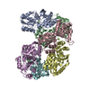
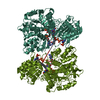
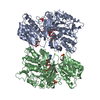
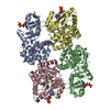
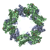
A: Hypothetical protein ybgI B: Hypothetical protein ybgI C: Hypothetical protein ybgI D: Hypothetical protein ybgI E: Hypothetical protein ybgI F: Hypothetical protein ybgI hetero molecules
The biological assembly is a hexamer generated by the application of the symmetry operations: -y, x-y, Z and y-x, -x, z. If applied to the entire asymmetric unit, these operations will generate 3 toroids (3 biological assemblies). If applied to any of the dimers, AB, CD or EF, these operations will produce one biological assembly.
-
Components
#1: Protein
HypotheticalproteinybgI / Hypothesis
Mass: 27106.980 Da / Num. of mol.: 6 Source method: isolated from a genetically manipulated source Source: (gene. exp.) Escherichia coli, Escherichia coli O157:H7 Genus: Escherichia, Escherichia / Species: , Escherichia coli / Strain: , O157:H7 / Gene: YBGI OR B0710 OR Z0861 OR ECS0735 / Plasmid: pDEST14 / Production host: Escherichia coli (E. coli) / Strain (production host): BL21 Star (DE3) / References: UniProt: P75743, UniProt: P0AFP6*PLUS
In the structure databanks used in Yorodumi, some data are registered as the other names, "COVID-19 virus" and "2019-nCoV". Here are the details of the virus and the list of structure data.
Jan 31, 2019. EMDB accession codes are about to change! (news from PDBe EMDB page)
EMDB accession codes are about to change! (news from PDBe EMDB page)
The allocation of 4 digits for EMDB accession codes will soon come to an end. Whilst these codes will remain in use, new EMDB accession codes will include an additional digit and will expand incrementally as the available range of codes is exhausted. The current 4-digit format prefixed with “EMD-” (i.e. EMD-XXXX) will advance to a 5-digit format (i.e. EMD-XXXXX), and so on. It is currently estimated that the 4-digit codes will be depleted around Spring 2019, at which point the 5-digit format will come into force.
The EM Navigator/Yorodumi systems omit the EMD- prefix.
Related info.:Q: What is EMD? / ID/Accession-code notation in Yorodumi/EM Navigator
Yorodumi is a browser for structure data from EMDB, PDB, SASBDB, etc.
This page is also the successor to EM Navigator detail page, and also detail information page/front-end page for Omokage search.
The word "yorodu" (or yorozu) is an old Japanese word meaning "ten thousand". "mi" (miru) is to see.
Related info.:EMDB / PDB / SASBDB / Comparison of 3 databanks / Yorodumi Search / Aug 31, 2016. New EM Navigator & Yorodumi / Yorodumi Papers / Jmol/JSmol / Function and homology information / Changes in new EM Navigator and Yorodumi
 Movie
Movie Controller


 Open data
 Basic information
 Components
 Hypothesis
  Keywords
 Function and homology information
 Authors
 Citation
 Structure visualization
 Downloads & links
 Other downloads
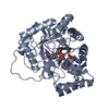


 PDBj
PDBj Assembly
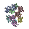


 Mass: 55.845 Da / Num. of mol.: 12 / Source method: obtained synthetically / Formula: Fe
 Mass: 18.015 Da / Num. of mol.: 637 / Source method: isolated from a natural source / Formula: H2O
 Sample preparation
 / Beamline: 22-ID / Wavelength: 0.9895,0.9793,0.9780
 Processing