 Movie
Movie Controller

[English] 日本語
 Yorodumi
- PDB-1nj2: Crystal structure of Prolyl-tRNA Synthetase from Methanothermobac... -
+ Open data

- Basic information
| Entry | Database: PDB / ID: 1nj2 | ||||||
|---|---|---|---|---|---|---|---|
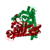
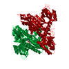
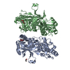
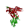
| Title | Crystal structure of Prolyl-tRNA Synthetase from Methanothermobacter thermautotrophicus | ||||||
 Components Components | Proline-tRNA Synthetase | ||||||
 Keywords Keywords |  LIGASE / Class-II tRNA synthetase LIGASE / Class-II tRNA synthetase | ||||||
| Function / homology |  Function and homology informationproline-tRNA ligase / proline-tRNA ligase activity / prolyl-tRNA aminoacylation / ATP binding / metal ion binding / cytoplasm Function and homology informationproline-tRNA ligase / proline-tRNA ligase activity / prolyl-tRNA aminoacylation / ATP binding / metal ion binding / cytoplasmSimilarity search - Function | ||||||
| Biological species |   Methanothermobacter thermautotrophicus (archaea) Methanothermobacter thermautotrophicus (archaea) | ||||||
| Method | X-RAY DIFFRACTION / SYNCHROTRON / MOLECULAR REPLACEMENT / Resolution: 3.11 Å | ||||||
 Authors Authors | Kamtekar, S. / Kennedy, W.D. / Wang, J. / Stathopoulos, C. / Soll, D. / Steitz, T.A. | ||||||
 Citation Citation | Journal: Proc.Natl.Acad.Sci.USA / Year: 2003 Title: The structural basis of cysteine aminoacylation of tRNAPro by prolyl-tRNA synthetases Authors: Kamtekar, S. / Kennedy, W.D. / Wang, J. / Stathopoulos, C. / Soll, D. / Steitz, T.A. | ||||||
| History |
|
- Structure visualization
| Structure viewer | Molecule: MolmilJmol/JSmol |
|---|
- Downloads & links
-Download
| PDBx/mmCIF format | 1nj2.cif.gz | 105.9 KB | Display | PDBx/mmCIF format |
|---|---|---|---|---|
| PDB format | pdb1nj2.ent.gz | 80.6 KB | Display | PDB format |
| PDBx/mmJSON format | 1nj2.json.gz | Tree view | PDBx/mmJSON format | |
| Others |  Other downloads Other downloads |
-Validation report
| Arichive directory | https://data.pdbj.org/pub/pdb/validation_reports/nj/1nj2ftp://data.pdbj.org/pub/pdb/validation_reports/nj/1nj2 | HTTPS FTP |
|---|
-Related structure data


-Links
 PDBj
PDBj
- Assembly
| Deposited unit | 
| ||||||||||
|---|---|---|---|---|---|---|---|---|---|---|---|
| 1 | 
| ||||||||||
| Unit cell |
| ||||||||||
| Details | The biological assembly is a dimer constructed by a twofold rotation. |
-Components
| #1: Protein | Mass: 58170.258 Da / Num. of mol.: 1 / Fragment: N terminally His Tagged Enzyme Source method: isolated from a genetically manipulated source Source: (gene. exp.) Methanothermobacter thermautotrophicus (archaea)Strain: Delta H Description: Strain supplemented with additional plasmid encoding rare tRNAs Gene: MTH611 / Plasmid: pET15b / Production host:  Escherichia coli (E. coli) / Strain (production host): BL21DE3 RecA- / References: UniProt: O26708, proline-tRNA ligase Escherichia coli (E. coli) / Strain (production host): BL21DE3 RecA- / References: UniProt: O26708, proline-tRNA ligase |
|---|---|
| #2: Chemical | ChemComp-ZN /   Mass: 65.409 Da / Num. of mol.: 1 / Source method: obtained synthetically / Formula: Zn Mass: 65.409 Da / Num. of mol.: 1 / Source method: obtained synthetically / Formula: Zn |
| #3: Chemical | ChemComp-MG /   Mass: 24.305 Da / Num. of mol.: 1 / Source method: obtained synthetically / Formula: Mg Mass: 24.305 Da / Num. of mol.: 1 / Source method: obtained synthetically / Formula: Mg |
| #4: Water | ChemComp-HOH / Water Mass: 18.015 Da / Num. of mol.: 13 / Source method: isolated from a natural source / Formula: H2O Mass: 18.015 Da / Num. of mol.: 13 / Source method: isolated from a natural source / Formula: H2O |
-Experimental details
-Experiment
| Experiment | Method: X-RAY DIFFRACTION / Number of used crystals: 1 |
|---|
- Sample preparation
| Crystal | Density Matthews: 4.98 Å3/Da / Density % sol: 75.13 % | |||||||||||||||||||||||||||||||||||||||||||||||||||||||||||||||
|---|---|---|---|---|---|---|---|---|---|---|---|---|---|---|---|---|---|---|---|---|---|---|---|---|---|---|---|---|---|---|---|---|---|---|---|---|---|---|---|---|---|---|---|---|---|---|---|---|---|---|---|---|---|---|---|---|---|---|---|---|---|---|---|---|
| Crystal grow | Temperature: 293 K / Method: vapor diffusion, hanging drop / pH: 7.5 Details: Tris, Beta-Mercaptoethanol, MgCl2,NaCl, pH 7.5, VAPOR DIFFUSION, HANGING DROP, temperature 293K | |||||||||||||||||||||||||||||||||||||||||||||||||||||||||||||||
| Crystal grow | *PLUS Temperature: 12-25 ℃ / pH: 8.5 / Method: vapor diffusion | |||||||||||||||||||||||||||||||||||||||||||||||||||||||||||||||
| Components of the solutions | *PLUS
|
-Data collection
| Diffraction | Mean temperature: 100 K |
|---|---|
| Diffraction source | Source: SYNCHROTRON / Site: CHESS  / Beamline: A1 / Wavelength: 0.945 Å / Beamline: A1 / Wavelength: 0.945 Å |
| Detector | Type: ADSC QUANTUM 4 / Detector: CCD / Date: Dec 20, 2000 |
| Radiation | Monochromator: Si(111) / Protocol: SINGLE WAVELENGTH / Monochromatic (M) / Laue (L): M / Scattering type: x-ray |
| Radiation wavelength | Wavelength: 0.945 Å / Relative weight: 1 |
| Reflection | Resolution: 3.1→30 Å / Num. all: 36062 / Num. obs: 35918 / % possible obs: 99.6 % / Observed criterion σ(I): -3 / Redundancy: 4.7 % / Rmerge(I) obs: 0.066 / Net I/σ(I): 22.8 |
| Reflection shell | Resolution: 3.1→3.21 Å / Rmerge(I) obs: 0.77 / Mean I/σ(I) obs: 2 / % possible all: 99.7 |
| Reflection | *PLUS Highest resolution: 3.1 Å |
| Reflection shell | *PLUS % possible obs: 99.7 % |
- Processing
| Software |
| ||||||||||||||||||||||||||||||||||||||||||||||||||||||||||||||||||||||||||||||||
|---|---|---|---|---|---|---|---|---|---|---|---|---|---|---|---|---|---|---|---|---|---|---|---|---|---|---|---|---|---|---|---|---|---|---|---|---|---|---|---|---|---|---|---|---|---|---|---|---|---|---|---|---|---|---|---|---|---|---|---|---|---|---|---|---|---|---|---|---|---|---|---|---|---|---|---|---|---|---|---|---|---|
| Refinement | Method to determine structure: MOLECULAR REPLACEMENT Starting model: Methanocaldococcus janaschii Proline tRNA Synthetase Resolution: 3.11→29.95 Å / Rfactor Rfree error: 0.005 / Isotropic thermal model: RESTRAINED / Cross valid method: THROUGHOUT / σ(F): 0 / Stereochemistry target values: Engh & Huber
| ||||||||||||||||||||||||||||||||||||||||||||||||||||||||||||||||||||||||||||||||
| Solvent computation | Solvent model: FLAT MODEL / Bsol: 24.3074 Å2 / ksol: 0.264032 e/Å3 | ||||||||||||||||||||||||||||||||||||||||||||||||||||||||||||||||||||||||||||||||
| Displacement parameters | Biso mean: 89.4 Å2
| ||||||||||||||||||||||||||||||||||||||||||||||||||||||||||||||||||||||||||||||||
| Refine analyze | Luzzati coordinate error free: 0.52 Å / Luzzati sigma a free: 0.75 Å | ||||||||||||||||||||||||||||||||||||||||||||||||||||||||||||||||||||||||||||||||
| Refinement step | Cycle: LAST / Resolution: 3.11→29.95 Å
| ||||||||||||||||||||||||||||||||||||||||||||||||||||||||||||||||||||||||||||||||
| Refine LS restraints |
| ||||||||||||||||||||||||||||||||||||||||||||||||||||||||||||||||||||||||||||||||
| LS refinement shell | Resolution: 3.11→3.29 Å / Rfactor Rfree error: 0.018 / Total num. of bins used: 6
| ||||||||||||||||||||||||||||||||||||||||||||||||||||||||||||||||||||||||||||||||
| Xplor file |
| ||||||||||||||||||||||||||||||||||||||||||||||||||||||||||||||||||||||||||||||||
| Refinement | *PLUS Highest resolution: 3.1 Å / Lowest resolution: 30 Å | ||||||||||||||||||||||||||||||||||||||||||||||||||||||||||||||||||||||||||||||||
| Solvent computation | *PLUS | ||||||||||||||||||||||||||||||||||||||||||||||||||||||||||||||||||||||||||||||||
| Displacement parameters | *PLUS | ||||||||||||||||||||||||||||||||||||||||||||||||||||||||||||||||||||||||||||||||
| Refine LS restraints | *PLUS
|
