 Movie
Movie Controller

[English] 日本語
 Yorodumi
- PDB-1ndh: CRYSTAL STRUCTURE OF NADH-CYTOCHROME B5 REDUCTASE FROM PIG LIVER ... -
+ Open data

- Basic information
| Entry | Database: PDB / ID: 1ndh | ||||||
|---|---|---|---|---|---|---|---|
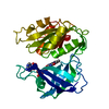
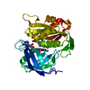
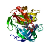
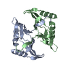
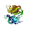
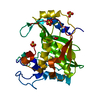
| Title | CRYSTAL STRUCTURE OF NADH-CYTOCHROME B5 REDUCTASE FROM PIG LIVER AT 2.4 ANGSTROMS RESOLUTION | ||||||
 Components Components | CYTOCHROME B5 REDUCTASE | ||||||
 Keywords Keywords | ELECTRON TRANSPORT (FLAVO PROTEIN) | ||||||
| Function / homology |  Function and homology informationcytochrome-b5 reductase / cytochrome-b5 reductase activity, acting on NAD(P)H / cholesterol biosynthetic process / FAD binding / flavin adenine dinucleotide binding / mitochondrial outer membrane / endoplasmic reticulum membrane / mitochondrion Function and homology informationcytochrome-b5 reductase / cytochrome-b5 reductase activity, acting on NAD(P)H / cholesterol biosynthetic process / FAD binding / flavin adenine dinucleotide binding / mitochondrial outer membrane / endoplasmic reticulum membrane / mitochondrionSimilarity search - Function | ||||||
| Biological species |  Sus scrofa (pig) Sus scrofa (pig) | ||||||
| Method | X-RAY DIFFRACTION / Resolution: 2.1 Å | ||||||
 Authors Authors | Nishida, H. / Miki, K. | ||||||
 Citation Citation | Journal: Biochemistry / Year: 1995 Title: Crystal structure of NADH-cytochrome b5 reductase from pig liver at 2.4 A resolution. Authors: Nishida, H. / Inaka, K. / Yamanaka, M. / Kaida, S. / Kobayashi, K. / Miki, K. #1: Journal: To be PublishedTitle: The Specific Arrangement of Three Amino Acidic Residues for Flavin Binding Barrel Structures in Nadh-Cytochrome B5 Reductase and the Other Flavin Dependent Reductases Authors: Nishida, H. / Inaka, K. / Miki, K. | ||||||
| History |
| ||||||
| Remark 700 | SHEET SHEET SHEET_ID: S1, BETA-BARREL. |
- Structure visualization
| Structure viewer | Molecule: MolmilJmol/JSmol |
|---|
- Downloads & links
-Download
| PDBx/mmCIF format | 1ndh.cif.gz | 69.4 KB | Display | PDBx/mmCIF format |
|---|---|---|---|---|
| PDB format | pdb1ndh.ent.gz | 50.3 KB | Display | PDB format |
| PDBx/mmJSON format | 1ndh.json.gz | Tree view | PDBx/mmJSON format | |
| Others |  Other downloads Other downloads |
-Validation report
| Arichive directory | https://data.pdbj.org/pub/pdb/validation_reports/nd/1ndhftp://data.pdbj.org/pub/pdb/validation_reports/nd/1ndh | HTTPS FTP |
|---|
-Related structure data
| Similar structure data |
|---|


-Links
 PDBj
PDBj

- Assembly
| Deposited unit | 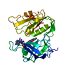
| ||||||||
|---|---|---|---|---|---|---|---|---|---|
| 1 |
| ||||||||
| Unit cell |
| ||||||||
| Atom site foot note | 1: CIS PROLINE - PRO 116 |
-Components
| #1: Protein | Mass: 30873.674 Da / Num. of mol.: 1 Source method: isolated from a genetically manipulated source Source: (gene. exp.) Sus scrofa (pig) / References: UniProt: P83686, cytochrome-b5 reductase |
|---|---|
| #2: Chemical | ChemComp-FAD / Flavin adenine dinucleotide  Mass: 785.550 Da / Num. of mol.: 1 / Source method: obtained synthetically / Formula: C27H33N9O15P2 / Comment: FAD*YM Mass: 785.550 Da / Num. of mol.: 1 / Source method: obtained synthetically / Formula: C27H33N9O15P2 / Comment: FAD*YM |
| #3: Water | ChemComp-HOH / Water Mass: 18.015 Da / Num. of mol.: 48 / Source method: isolated from a natural source / Formula: H2O Mass: 18.015 Da / Num. of mol.: 48 / Source method: isolated from a natural source / Formula: H2O |
-Experimental details
-Experiment
| Experiment | Method: X-RAY DIFFRACTION |
|---|
- Sample preparation
| Crystal | Density Matthews: 2.51 Å3/Da / Density % sol: 51.06 % | |||||||||||||||||||||||||||||||||||
|---|---|---|---|---|---|---|---|---|---|---|---|---|---|---|---|---|---|---|---|---|---|---|---|---|---|---|---|---|---|---|---|---|---|---|---|---|
| Crystal grow | *PLUS Temperature: 4 or 20 ℃ / Method: vapor diffusion, sitting drop / Details: Miki, K., (1987) J. Biol. Chem., 262, 11801. / PH range low: 8 / PH range high: 6.8 | |||||||||||||||||||||||||||||||||||
| Components of the solutions | *PLUS
|
-Data collection
| Radiation | Scattering type: x-ray |
|---|---|
| Radiation wavelength | Relative weight: 1 |
| Reflection | *PLUS Num. obs: 18695 / Rmerge(I) obs: 0.055 |
- Processing
| Software |
| ||||||||||||||||||||||||||||||||||||||||||||||||||||||||||||
|---|---|---|---|---|---|---|---|---|---|---|---|---|---|---|---|---|---|---|---|---|---|---|---|---|---|---|---|---|---|---|---|---|---|---|---|---|---|---|---|---|---|---|---|---|---|---|---|---|---|---|---|---|---|---|---|---|---|---|---|---|---|
| Refinement | Resolution: 2.1→5 Å /
| ||||||||||||||||||||||||||||||||||||||||||||||||||||||||||||
| Refinement step | Cycle: LAST / Resolution: 2.1→5 Å
| ||||||||||||||||||||||||||||||||||||||||||||||||||||||||||||
| Refine LS restraints |
| ||||||||||||||||||||||||||||||||||||||||||||||||||||||||||||
| Refinement | *PLUS Highest resolution: 2.1 Å / Num. reflection obs: 10350 / Rfactor Rwork: 0.237 | ||||||||||||||||||||||||||||||||||||||||||||||||||||||||||||
| Solvent computation | *PLUS | ||||||||||||||||||||||||||||||||||||||||||||||||||||||||||||
| Displacement parameters | *PLUS | ||||||||||||||||||||||||||||||||||||||||||||||||||||||||||||
| Refine LS restraints | *PLUS Type: x_angle_d / Dev ideal: 3.3 |
