 Movie
Movie Controller

[English] 日本語
 Yorodumi
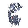
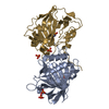
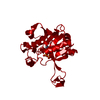
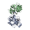
- PDB-1n4o: Crystal structure of the Class A beta-lactamase L2 from Stenotrop... -
+ Open data

- Basic information
| Entry | Database: PDB / ID: 1n4o | ||||||
|---|---|---|---|---|---|---|---|
| Title | Crystal structure of the Class A beta-lactamase L2 from Stenotrophomonas maltophilia | ||||||
 Components Components | L2 beta-lactamase | ||||||
 Keywords Keywords |  HYDROLASE / BETA-LACTAMASE / CLASS A / L2 HYDROLASE / BETA-LACTAMASE / CLASS A / L2 | ||||||
| Function / homology |  Function and homology information Function and homology informationbeta-lactam antibiotic catabolic process / beta-lactamase activity / beta-lactamase / response to antibioticSimilarity search - Function | ||||||
| Biological species |  Stenotrophomonas maltophilia (bacteria) Stenotrophomonas maltophilia (bacteria) | ||||||
| Method | X-RAY DIFFRACTION / SYNCHROTRON / MOLECULAR REPLACEMENT / Resolution: 1.85 Å | ||||||
 Authors Authors | Pernot, L. / Petrella, S. / Sougakoff, W. | ||||||
 Citation Citation | Journal: To be Published Title: Role of the disulfide bridge Cys69-Cys238 in class A b-lactamases : a structural and biochemical investigation on the b-lactamase L2 from Stenotrophomonas maltophilia Authors: Pernot, L. / Petrella, S. / Sougakoff, W. | ||||||
| History |
| ||||||
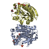
| Remark 300 | BIOMOLECULE: 1 The asymetric unit contains a homodimer generated by a 2-fold non-crystallographic ...BIOMOLECULE: 1 The asymetric unit contains a homodimer generated by a 2-fold non-crystallographic symmetry axis. The transformation applied to chain A gives chain B. Rotation : -0.87108099 0.47216475 0.13519730 0.47738487 0.74928564 0.45899314 0.11541899 0.46436137 -0.87809283 Translation : 37.0187 -14.5258 17.0446 |
- Structure visualization
| Structure viewer | Molecule: MolmilJmol/JSmol |
|---|
- Downloads & links
-Download
| PDBx/mmCIF format | 1n4o.cif.gz | 119.2 KB | Display | PDBx/mmCIF format |
|---|---|---|---|---|
| PDB format | pdb1n4o.ent.gz | 92.3 KB | Display | PDB format |
| PDBx/mmJSON format | 1n4o.json.gz | Tree view | PDBx/mmJSON format | |
| Others |  Other downloads Other downloads |
-Validation report
| Arichive directory | https://data.pdbj.org/pub/pdb/validation_reports/n4/1n4oftp://data.pdbj.org/pub/pdb/validation_reports/n4/1n4o | HTTPS FTP |
|---|
-Related structure data
| Related structure data |  1bzaS S: Starting model for refinement |
|---|---|
| Similar structure data |


-Links
 PDBj
PDBj


- Assembly
| Deposited unit | 
| ||||||||
|---|---|---|---|---|---|---|---|---|---|
| 1 |
| ||||||||
| Unit cell |
| ||||||||
| Details | The asymetric unit contains a homodimer generated by a 2-fold non-crystallographic symmetry axis. The transformation applied to chain A gives chain B. Rotation : -0.87108099 0.47216475 0.13519730 0.47738487 0.74928564 0.45899314 0.11541899 0.46436137 -0.87809283 Translation : 37.0187 -14.5258 17.0446 |
-Components
| #1: Protein | Mass: 29164.059 Da / Num. of mol.: 2 Source method: isolated from a genetically manipulated source Source: (gene. exp.) Stenotrophomonas maltophilia (bacteria)Strain: 405 / Gene: blaL2 / Plasmid: pET29(a+) / Species (production host): Escherichia coli / Production host: Escherichia coli BL21 (bacteria) / Strain (production host): BL21 / References: UniProt: Q9RBQ1, beta-lactamase#2: Chemical | ChemComp-SO4 / Sulfate  Mass: 96.063 Da / Num. of mol.: 4 / Source method: obtained synthetically / Formula: SO4 Mass: 96.063 Da / Num. of mol.: 4 / Source method: obtained synthetically / Formula: SO4#3: Water | ChemComp-HOH / | Water Mass: 18.015 Da / Num. of mol.: 315 / Source method: isolated from a natural source / Formula: H2O Mass: 18.015 Da / Num. of mol.: 315 / Source method: isolated from a natural source / Formula: H2O |
|---|
-Experimental details
-Experiment
| Experiment | Method: X-RAY DIFFRACTION / Number of used crystals: 1 |
|---|
- Sample preparation
| Crystal | Density Matthews: 3.55 Å3/Da / Density % sol: 65.05 % |
|---|---|
| Crystal grow | Temperature: 291 K / Method: vapor diffusion, hanging drop / pH: 6.5 Details: ammonium sulfate 1.5M, sodium cacodylate 0.1M, pH 6.5, VAPOR DIFFUSION, HANGING DROP, temperature 291K |
-Data collection
| Diffraction | Mean temperature: 283 K |
|---|---|
| Diffraction source | Source: SYNCHROTRON / Site: LURE  / Beamline: DW32 / Wavelength: 0.9485 Å / Beamline: DW32 / Wavelength: 0.9485 Å |
| Detector | Type: MARRESEARCH / Detector: IMAGE PLATE / Date: May 25, 2001 |
| Radiation | Monochromator: Si (1 1 1) / Protocol: SINGLE WAVELENGTH / Monochromatic (M) / Laue (L): M / Scattering type: x-ray |
| Radiation wavelength | Wavelength: 0.9485 Å / Relative weight: 1 |
| Reflection | Resolution: 1.85→30 Å / Num. obs: 75048 / % possible obs: 99.2 % / Observed criterion σ(F): 0 / Observed criterion σ(I): 2.5 / Redundancy: 6.1 % / Biso Wilson estimate: 25.4 Å2 / Rmerge(I) obs: 0.064 / Net I/σ(I): 22.7 |
| Reflection shell | Resolution: 1.85→1.95 Å / Redundancy: 5.5 % / Rmerge(I) obs: 0.286 / Mean I/σ(I) obs: 2.6 / Num. unique all: 10357 / % possible all: 95.2 |
- Processing
| Software |
| |||||||||||||||||||||||||||||||||||||||||||||||||||||||||||||||||||||||||||
|---|---|---|---|---|---|---|---|---|---|---|---|---|---|---|---|---|---|---|---|---|---|---|---|---|---|---|---|---|---|---|---|---|---|---|---|---|---|---|---|---|---|---|---|---|---|---|---|---|---|---|---|---|---|---|---|---|---|---|---|---|---|---|---|---|---|---|---|---|---|---|---|---|---|---|---|---|
| Refinement | Method to determine structure: MOLECULAR REPLACEMENT Starting model: PDB ENTRY 1BZA Resolution: 1.85→30 Å / Cor.coef. Fo:Fc: 0.968 / Cor.coef. Fo:Fc free: 0.961 / SU B: 1.669 / SU ML: 0.051 / Isotropic thermal model: ISOTROPIC / Cross valid method: THROUGHOUT / σ(F): 0 / σ(I): 0 / ESU R: 0.08 / ESU R Free: 0.078 / Stereochemistry target values: Engh & Huber
| |||||||||||||||||||||||||||||||||||||||||||||||||||||||||||||||||||||||||||
| Solvent computation | Ion probe radii: 0.8 Å / Shrinkage radii: 0.8 Å / VDW probe radii: 1.4 Å / Solvent model: BABINET MODEL WITH MASK | |||||||||||||||||||||||||||||||||||||||||||||||||||||||||||||||||||||||||||
| Displacement parameters | Biso mean: 17.3 Å2 | |||||||||||||||||||||||||||||||||||||||||||||||||||||||||||||||||||||||||||
| Refine analyze |
| |||||||||||||||||||||||||||||||||||||||||||||||||||||||||||||||||||||||||||
| Refinement step | Cycle: LAST / Resolution: 1.85→30 Å
| |||||||||||||||||||||||||||||||||||||||||||||||||||||||||||||||||||||||||||
| Refine LS restraints |
| |||||||||||||||||||||||||||||||||||||||||||||||||||||||||||||||||||||||||||
| LS refinement shell | Resolution: 1.85→1.97 Å / Rfactor Rfree error: 0.007
|
