 Movie
Movie Controller

[English] 日本語
 Yorodumi
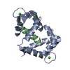
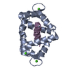
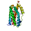
- PDB-1mxe: Structure of the Complex of Calmodulin with the Target Sequence o... -
+ Open data

- Basic information
| Entry | Database: PDB / ID: 1mxe | ||||||
|---|---|---|---|---|---|---|---|
| Title | Structure of the Complex of Calmodulin with the Target Sequence of CaMKI | ||||||
 Components Components |
| ||||||
 Keywords Keywords | METAL BINDING PROTEIN / Calmodulin-Pepide Complex /  Calmodulin / CaMKI / XRAY Calmodulin / CaMKI / XRAY | ||||||
| Function / homology |  Function and homology information Function and homology informationActivation of RAC1 downstream of NMDARs / positive regulation of syncytium formation by plasma membrane fusion / metarhodopsin inactivation / myosin VI complex / myosin VI head/neck binding / myosin VII complex / photoreceptor cell axon guidance / negative regulation of opsin-mediated signaling pathway / rhabdomere / rhabdomere development ...Activation of RAC1 downstream of NMDARs / positive regulation of syncytium formation by plasma membrane fusion / metarhodopsin inactivation / myosin VI complex / myosin VI head/neck binding / myosin VII complex / photoreceptor cell axon guidance / negative regulation of opsin-mediated signaling pathway / rhabdomere / rhabdomere development / myosin V complex / : / kinetochore organization / positive regulation of synapse structural plasticity / regulation of muscle cell differentiation / : / actin filament-based movement / Ca2+/calmodulin-dependent protein kinase / rhodopsin mediated signaling pathway / Neutrophil degranulation / regulation of protein binding / myosin V binding / channel regulator activity / cellular response to ethanol / calmodulin-dependent protein kinase activity / nucleocytoplasmic transport / positive regulation of dendritic spine development / muscle cell cellular homeostasis / regulation of synapse organization / myosin heavy chain binding / positive regulation of muscle cell differentiation / mitotic spindle pole / centriole replication / enzyme regulator activity / centriole / positive regulation of protein export from nucleus / sensory perception of sound / positive regulation of protein serine/threonine kinase activity / positive regulation of neuron projection development / spindle / mitotic spindle / regulation of protein localization / sensory perception of smell / nervous system development / cell cortex / midbody / postsynaptic density / cell differentiation / calmodulin binding / intracellular signal transduction / cell cycle / protein phosphorylation / protein serine kinase activity / centrosome / glutamatergic synapse / calcium ion binding / signal transduction / positive regulation of transcription by RNA polymerase II / nucleoplasm / ATP binding / nucleus / cytosol / cytoplasmSimilarity search - Function | ||||||
| Biological species |  Drosophila melanogaster (fruit fly) Drosophila melanogaster (fruit fly) | ||||||
| Method | X-RAY DIFFRACTION / SYNCHROTRON / MAD / Resolution: 1.7 Å | ||||||
 Authors Authors | Clapperton, J.A. / Martin, S.R. / Smerdon, S.J. / Gamblin, S.J. / Bayley, P.M. | ||||||
 Citation Citation | Journal: Biochemistry / Year: 2002 Title: Structure of the Complex of Calmodulin with the Target Sequence of Calmodulin-Dependent Protein Kinase I: Studies of the Kinase Activation Mechanism Authors: Clapperton, J.A. / Martin, S.R. / Smerdon, S.J. / Gamblin, S.J. / Bayley, P.M. | ||||||
| History |
|
- Structure visualization
| Structure viewer | Molecule: MolmilJmol/JSmol |
|---|
- Downloads & links
-Download
| PDBx/mmCIF format | 1mxe.cif.gz | 154.4 KB | Display | PDBx/mmCIF format |
|---|---|---|---|---|
| PDB format | pdb1mxe.ent.gz | 122.9 KB | Display | PDB format |
| PDBx/mmJSON format | 1mxe.json.gz | Tree view | PDBx/mmJSON format | |
| Others |  Other downloads Other downloads |
-Validation report
| Arichive directory | https://data.pdbj.org/pub/pdb/validation_reports/mx/1mxeftp://data.pdbj.org/pub/pdb/validation_reports/mx/1mxe | HTTPS FTP |
|---|
-Related structure data
| Similar structure data |
|---|


-Links
 PDBj
PDBj

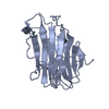
- Assembly
| Deposited unit | 
| ||||||||
|---|---|---|---|---|---|---|---|---|---|
| 1 | 
| ||||||||
| 2 | 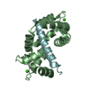
| ||||||||
| Unit cell |
|
-Components
| #1: Protein | Mass: 16694.324 Da / Num. of mol.: 2 Source method: isolated from a genetically manipulated source Source: (gene. exp.) Drosophila melanogaster (fruit fly) / Species (production host): Escherichia coli / Production host:  Escherichia coli BL21 (bacteria) / Strain (production host): BL21 / References: UniProt: P62152 Escherichia coli BL21 (bacteria) / Strain (production host): BL21 / References: UniProt: P62152#2: Protein/peptide | Mass: 2997.628 Da / Num. of mol.: 2 / Fragment: Calmodulin Binding Domain / Source method: obtained synthetically / Details: Peptide synthesis / References: UniProt: Q63450, EC: 2.7.1.123 #3: Chemical | ChemComp-CA /   Mass: 40.078 Da / Num. of mol.: 8 / Source method: obtained synthetically / Formula: Ca Mass: 40.078 Da / Num. of mol.: 8 / Source method: obtained synthetically / Formula: Ca#4: Water | ChemComp-HOH / | Water Mass: 18.015 Da / Num. of mol.: 234 / Source method: isolated from a natural source / Formula: H2O Mass: 18.015 Da / Num. of mol.: 234 / Source method: isolated from a natural source / Formula: H2O |
|---|
-Experimental details
-Experiment
| Experiment | Method: X-RAY DIFFRACTION / Number of used crystals: 1 |
|---|
- Sample preparation
| Crystal | Density Matthews: 2.11 Å3/Da / Density % sol: 41.12 % | ||||||||||||||||||||||||||||||||||||||||||||||||||||||||
|---|---|---|---|---|---|---|---|---|---|---|---|---|---|---|---|---|---|---|---|---|---|---|---|---|---|---|---|---|---|---|---|---|---|---|---|---|---|---|---|---|---|---|---|---|---|---|---|---|---|---|---|---|---|---|---|---|---|
| Crystal grow | Temperature: 289 K / Method: vapor diffusion, hanging drop / pH: 5.5 Details: Cacodylate, Calcium chloride, MPD, pH 5.5, VAPOR DIFFUSION, HANGING DROP, temperature 289K | ||||||||||||||||||||||||||||||||||||||||||||||||||||||||
| Crystal grow | *PLUS Temperature: 16 ℃ / pH: 7.8 | ||||||||||||||||||||||||||||||||||||||||||||||||||||||||
| Components of the solutions | *PLUS
|
-Data collection
| Diffraction | Mean temperature: 100 K | |||||||||||||||
|---|---|---|---|---|---|---|---|---|---|---|---|---|---|---|---|---|
| Diffraction source | Source: SYNCHROTRON / Site: ESRF  / Beamline: ID14-1 / Wavelength: 0.934, 0.9500, 0.9791, 0.9794 / Beamline: ID14-1 / Wavelength: 0.934, 0.9500, 0.9791, 0.9794 | |||||||||||||||
| Detector | Type: ADSC QUANTUM 4 / Detector: CCD / Date: Jun 9, 2002 | |||||||||||||||
| Radiation | Protocol: MAD / Monochromatic (M) / Laue (L): M / Scattering type: x-ray | |||||||||||||||
| Radiation wavelength |
| |||||||||||||||
| Reflection | Resolution: 1.7→30 Å / Num. all: 39543 / Num. obs: 38674 / % possible obs: 99.9 % / Observed criterion σ(F): 2 / Observed criterion σ(I): 2 / Rmerge(I) obs: 0.077 | |||||||||||||||
| Reflection shell | Resolution: 1.7→1.76 Å / Rmerge(I) obs: 0.217 / % possible all: 100 | |||||||||||||||
| Reflection | *PLUS Redundancy: 23.1 % | |||||||||||||||
| Reflection shell | *PLUS % possible obs: 100 % / Mean I/σ(I) obs: 7 |
- Processing
| Software |
| |||||||||||||||||||||||||||||||||||||||||||||||||||||||||||||||||||||||||||||||||||||||||||||||||||||||||||||||||||||||||||||||||||||||||||||||||||||||||||||||||||||||||||||||
|---|---|---|---|---|---|---|---|---|---|---|---|---|---|---|---|---|---|---|---|---|---|---|---|---|---|---|---|---|---|---|---|---|---|---|---|---|---|---|---|---|---|---|---|---|---|---|---|---|---|---|---|---|---|---|---|---|---|---|---|---|---|---|---|---|---|---|---|---|---|---|---|---|---|---|---|---|---|---|---|---|---|---|---|---|---|---|---|---|---|---|---|---|---|---|---|---|---|---|---|---|---|---|---|---|---|---|---|---|---|---|---|---|---|---|---|---|---|---|---|---|---|---|---|---|---|---|---|---|---|---|---|---|---|---|---|---|---|---|---|---|---|---|---|---|---|---|---|---|---|---|---|---|---|---|---|---|---|---|---|---|---|---|---|---|---|---|---|---|---|---|---|---|---|---|---|---|
| Refinement | Method to determine structure: MAD / Resolution: 1.7→29.75 Å / Cor.coef. Fo:Fc: 0.96 / Cor.coef. Fo:Fc free: 0.943 / SU B: 5.168 / SU ML: 0.072 / Cross valid method: THROUGHOUT / σ(F): 0 / ESU R: 0.173 / ESU R Free: 0.112 / Details: HYDROGENS HAVE BEEN ADDED IN THE RIDING POSITIONS
| |||||||||||||||||||||||||||||||||||||||||||||||||||||||||||||||||||||||||||||||||||||||||||||||||||||||||||||||||||||||||||||||||||||||||||||||||||||||||||||||||||||||||||||||
| Solvent computation | Ion probe radii: 0.8 Å / Shrinkage radii: 0.8 Å / VDW probe radii: 1.4 Å / Solvent model: BABINET MODEL WITH MASK | |||||||||||||||||||||||||||||||||||||||||||||||||||||||||||||||||||||||||||||||||||||||||||||||||||||||||||||||||||||||||||||||||||||||||||||||||||||||||||||||||||||||||||||||
| Displacement parameters | Biso mean: 18.529 Å2
| |||||||||||||||||||||||||||||||||||||||||||||||||||||||||||||||||||||||||||||||||||||||||||||||||||||||||||||||||||||||||||||||||||||||||||||||||||||||||||||||||||||||||||||||
| Refinement step | Cycle: LAST / Resolution: 1.7→29.75 Å
| |||||||||||||||||||||||||||||||||||||||||||||||||||||||||||||||||||||||||||||||||||||||||||||||||||||||||||||||||||||||||||||||||||||||||||||||||||||||||||||||||||||||||||||||
| Refine LS restraints |
| |||||||||||||||||||||||||||||||||||||||||||||||||||||||||||||||||||||||||||||||||||||||||||||||||||||||||||||||||||||||||||||||||||||||||||||||||||||||||||||||||||||||||||||||
| LS refinement shell | Resolution: 1.7→1.744 Å / Total num. of bins used: 20 /
| |||||||||||||||||||||||||||||||||||||||||||||||||||||||||||||||||||||||||||||||||||||||||||||||||||||||||||||||||||||||||||||||||||||||||||||||||||||||||||||||||||||||||||||||
| Refinement TLS params. | Method: refined / Refine-ID: X-RAY DIFFRACTION
| |||||||||||||||||||||||||||||||||||||||||||||||||||||||||||||||||||||||||||||||||||||||||||||||||||||||||||||||||||||||||||||||||||||||||||||||||||||||||||||||||||||||||||||||
| Refinement TLS group |
| |||||||||||||||||||||||||||||||||||||||||||||||||||||||||||||||||||||||||||||||||||||||||||||||||||||||||||||||||||||||||||||||||||||||||||||||||||||||||||||||||||||||||||||||
| Refinement | *PLUS Lowest resolution: 30 Å / % reflection Rfree: 5 % / Rfactor Rfree: 0.228 / Rfactor Rwork: 0.188 | |||||||||||||||||||||||||||||||||||||||||||||||||||||||||||||||||||||||||||||||||||||||||||||||||||||||||||||||||||||||||||||||||||||||||||||||||||||||||||||||||||||||||||||||
| Solvent computation | *PLUS | |||||||||||||||||||||||||||||||||||||||||||||||||||||||||||||||||||||||||||||||||||||||||||||||||||||||||||||||||||||||||||||||||||||||||||||||||||||||||||||||||||||||||||||||
| Displacement parameters | *PLUS | |||||||||||||||||||||||||||||||||||||||||||||||||||||||||||||||||||||||||||||||||||||||||||||||||||||||||||||||||||||||||||||||||||||||||||||||||||||||||||||||||||||||||||||||
| Refine LS restraints | *PLUS
| |||||||||||||||||||||||||||||||||||||||||||||||||||||||||||||||||||||||||||||||||||||||||||||||||||||||||||||||||||||||||||||||||||||||||||||||||||||||||||||||||||||||||||||||
| LS refinement shell | *PLUS |
