 Movie
Movie Controller

[English] 日本語
 Yorodumi
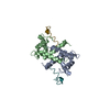
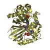
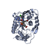
- PDB-1mwn: Solution NMR structure of S100B bound to the high-affinity target... -
+ Open data

- Basic information
| Entry | Database: PDB / ID: 1mwn | ||||||
|---|---|---|---|---|---|---|---|
| Title | Solution NMR structure of S100B bound to the high-affinity target peptide TRTK-12 | ||||||
 Components Components |
| ||||||
 Keywords Keywords |  STRUCTURAL PROTEIN / S100B / TRTK-12 / Calcium-binding / EF-hand / S100 protein / four helix bundle / helix loop helix / protein-peptide complex / 20 structures STRUCTURAL PROTEIN / S100B / TRTK-12 / Calcium-binding / EF-hand / S100 protein / four helix bundle / helix loop helix / protein-peptide complex / 20 structures | ||||||
| Function / homology |  Function and homology information Function and homology informationnegative regulation of skeletal muscle cell differentiation / TRAF6 mediated NF-kB activation / Advanced glycosylation endproduct receptor signaling / TAK1-dependent IKK and NF-kappa-B activation / WASH complex / F-actin capping protein complex / adaptive thermogenesis / sympathetic neuron projection extension / positive regulation of myelination / RAGE receptor binding ...negative regulation of skeletal muscle cell differentiation / TRAF6 mediated NF-kB activation / Advanced glycosylation endproduct receptor signaling / TAK1-dependent IKK and NF-kappa-B activation / WASH complex / F-actin capping protein complex / adaptive thermogenesis / sympathetic neuron projection extension / positive regulation of myelination / RAGE receptor binding / COPI-independent Golgi-to-ER retrograde traffic / cell junction assembly / barbed-end actin filament capping / response to methylmercury / ion binding / astrocyte differentiation / S100 protein binding / Sensory processing of sound by inner hair cells of the cochlea / neuron projection extension / regulation of neuronal synaptic plasticity / Advanced glycosylation endproduct receptor signaling / COPI-mediated anterograde transport / positive regulation of synaptic transmission / response to glucocorticoid / ruffle / HSP90 chaperone cycle for steroid hormone receptors (SHR) in the presence of ligand / MHC class II antigen presentation / positive regulation of neuron differentiation / Gene and protein expression by JAK-STAT signaling after Interleukin-12 stimulation / long-term synaptic potentiation / tau protein binding / memory / calcium-dependent protein binding / actin filament binding / actin cytoskeleton / actin binding / Factors involved in megakaryocyte development and platelet production / regulation of cell shape / cellular response to hypoxia / actin cytoskeleton organization / protein-containing complex assembly / positive regulation of canonical NF-kappaB signal transduction / learning or memory / cytoskeleton / cell adhesion / cadherin binding / positive regulation of apoptotic process / signaling receptor binding / neuronal cell body / calcium ion binding / positive regulation of cell population proliferation / perinuclear region of cytoplasm / protein homodimerization activity / extracellular space / extracellular exosome / zinc ion binding / extracellular region / identical protein binding / nucleus / cytosol / cytoplasmSimilarity search - Function | ||||||
| Biological species |  Rattus norvegicus (Norway rat) Rattus norvegicus (Norway rat) | ||||||
| Method | SOLUTION NMR / distance geometry, simulated annealing | ||||||
 Authors Authors | Inman, K.G. / Yang, R. / Rustandi, R.R. / Miller, K.E. / Baldisseri, D.M. / Weber, D.J. | ||||||
 Citation Citation | Journal: J.Mol.Biol. / Year: 2002 Title: Solution NMR structure of S100B bound to the high-affinity target peptide TRTK-12 Authors: Inman, K.G. / Yang, R. / Rustandi, R.R. / Miller, K.E. / Baldisseri, D.M. / Weber, D.J. #1: Journal: Nat.Struct.Biol. / Year: 2000Title: Structure of the negative regulatory domain of p53 bound to S100B(betabeta) Authors: Rustandi, R.R. / Baldisseri, D.M. / Weber, D.J. #2: Journal: Biochemistry / Year: 1998Title: Solution structure of calcium-bound rat S100B(betabeta) as determined by nuclear magnetic resonance spectroscopy Authors: Drohat, A.C. / Baldisseri, D.M. / Rustandi, R.R. / Weber, D.J. #3: Journal: Biochemistry / Year: 1996Title: Solution structure of rat apo-S100B(betabeta) as determined by NMR spectroscopy Authors: Drohat, A.C. / Amburgey, J.C. / Abildgaard, F. / Starich, M.R. / Baldisseri, D.M. / Weber, D.J. | ||||||
| History |
|
- Structure visualization
| Structure viewer | Molecule: MolmilJmol/JSmol |
|---|
- Downloads & links
-Download
| PDBx/mmCIF format | 1mwn.cif.gz | 1.3 MB | Display | PDBx/mmCIF format |
|---|---|---|---|---|
| PDB format | pdb1mwn.ent.gz | 1.1 MB | Display | PDB format |
| PDBx/mmJSON format | 1mwn.json.gz | Tree view | PDBx/mmJSON format | |
| Others |  Other downloads Other downloads |
-Validation report
| Arichive directory | https://data.pdbj.org/pub/pdb/validation_reports/mw/1mwnftp://data.pdbj.org/pub/pdb/validation_reports/mw/1mwn | HTTPS FTP |
|---|
-Related structure data
| Related structure data | |
|---|---|
| Similar structure data |


-Links
 PDBj
PDBj


- Assembly
| Deposited unit | 
| |||||||||
|---|---|---|---|---|---|---|---|---|---|---|
| 1 |
| |||||||||
| NMR ensembles |
|
-Components
| #1: Protein | Mass: 10758.048 Da / Num. of mol.: 2 Source method: isolated from a genetically manipulated source Source: (gene. exp.) Rattus norvegicus (Norway rat) / Gene: S100B / Plasmid: pET11b / Production host:  Escherichia coli (E. coli) / Strain (production host): HMS174 (DE3) / References: UniProt: P04631 Escherichia coli (E. coli) / Strain (production host): HMS174 (DE3) / References: UniProt: P04631#2: Protein/peptide | Mass: 1477.728 Da / Num. of mol.: 2 / Fragment: Residues 265-276 / Source method: obtained synthetically Details: The peptide was chemically synthesized. The sequence of the peptide is naturally found in Homo sapiens (human). References: UniProt: P52907 #3: Chemical | ChemComp-CA /   Mass: 40.078 Da / Num. of mol.: 4 / Source method: obtained synthetically / Formula: Ca Mass: 40.078 Da / Num. of mol.: 4 / Source method: obtained synthetically / Formula: Ca |
|---|
-Experimental details
-Experiment
| Experiment | Method: SOLUTION NMR | ||||||||||||||||||||||||||||||||
|---|---|---|---|---|---|---|---|---|---|---|---|---|---|---|---|---|---|---|---|---|---|---|---|---|---|---|---|---|---|---|---|---|---|
| NMR experiment |
| ||||||||||||||||||||||||||||||||
| NMR details | Text: This structure was determined using triple-resonance NMR spectroscopy. |
- Sample preparation
| Details | Contents: 2.2 mM S100B (subunit concentration), 5.2 mM CaCl2, 2.6 mM TRTK-12 peptide, 10 mM tris-d11, 15 mM NaCl, 0.1 mM EDTA, 5 mM DTT, 0.35 mM NaN3 Solvent system: 95% H2O/5% D2O |
|---|---|
| Sample conditions | Ionic strength: 25 mM / pH: 6.5 / Pressure: ambient / Temperature: 310 K |
| Crystal grow | *PLUS Method: other / Details: NMR |
-NMR measurement
| Radiation | Protocol: SINGLE WAVELENGTH / Monochromatic (M) / Laue (L): M | |||||||||||||||
|---|---|---|---|---|---|---|---|---|---|---|---|---|---|---|---|---|
| Radiation wavelength | Relative weight: 1 | |||||||||||||||
| NMR spectrometer |
|
- Processing
| NMR software |
| ||||||||||||||||
|---|---|---|---|---|---|---|---|---|---|---|---|---|---|---|---|---|---|
| Refinement | Method: distance geometry, simulated annealing / Software ordinal: 1 | ||||||||||||||||
| NMR representative | Selection criteria: closest to the average | ||||||||||||||||
| NMR ensemble | Conformer selection criteria: structures with the lowest energy Conformers calculated total number: 200 / Conformers submitted total number: 20 |
