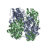
 Movie
Movie Controller


+ Open data

- Basic information
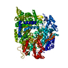
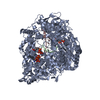
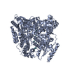
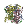
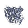
| Entry | Database: PDB / ID: 1mwh | ||||||
|---|---|---|---|---|---|---|---|
| Title | REOVIRUS POLYMERASE LAMBDA3 BOUND TO MRNA CAP ANALOG | ||||||
 Components Components | MINOR CORE PROTEIN LAMBDA 3 | ||||||
 Keywords Keywords |  VIRAL PROTEIN / polymerase / polymerase-cap analog complex / right hand configuration VIRAL PROTEIN / polymerase / polymerase-cap analog complex / right hand configuration | ||||||
| Function / homology |  Function and homology information Function and homology informationviral genome replication / viral nucleocapsid / hydrolase activity / RNA-directed RNA polymerase / RNA-dependent RNA polymerase activity / nucleotide binding / RNA bindingSimilarity search - Function | ||||||
| Biological species |  Reovirus sp. Reovirus sp. | ||||||
| Method | X-RAY DIFFRACTION / SYNCHROTRON / MOLECULAR REPLACEMENT / Resolution: 2.5 Å | ||||||
 Authors Authors | Tao, Y. / Farsetta, D.L. / Nibert, M.L. / Harrison, S.C. | ||||||
 Citation Citation | Journal: Cell(Cambridge,Mass.) / Year: 2002 Title: RNA Synthesis in a Cage-Structural Studies of Reovirus Polymerase lambda3 Authors: Tao, Y. / Farsetta, D.L. / Nibert, M.L. / Harrison, S.C. | ||||||
| History |
|
- Structure visualization
| Structure viewer | Molecule: MolmilJmol/JSmol |
|---|
- Downloads & links
-Download
| PDBx/mmCIF format | 1mwh.cif.gz | 276.4 KB | Display | PDBx/mmCIF format |
|---|---|---|---|---|
| PDB format | pdb1mwh.ent.gz | 218.4 KB | Display | PDB format |
| PDBx/mmJSON format | 1mwh.json.gz | Tree view | PDBx/mmJSON format | |
| Others |  Other downloads Other downloads |
-Validation report
| Arichive directory | https://data.pdbj.org/pub/pdb/validation_reports/mw/1mwhftp://data.pdbj.org/pub/pdb/validation_reports/mw/1mwh | HTTPS FTP |
|---|
-Related structure data
| Related structure data |  1mukSC  1n1hC  1n35C  1n38C S: Starting model for refinement C: citing same article ( |
|---|---|
| Similar structure data |

-Links
 PDBj
PDBj

- Assembly
| Deposited unit | 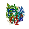
| ||||||||
|---|---|---|---|---|---|---|---|---|---|
| 1 |
| ||||||||
| Unit cell |
|
-Components
| #1: Protein | Mass: 142423.797 Da / Num. of mol.: 1 Source method: isolated from a genetically manipulated source Source: (gene. exp.) Reovirus sp. / Gene: L1 / Cell line (production host): SF21 / Production host:   Spodoptera frugiperda (fall armyworm) / References: UniProt: P17378, UniProt: P0CK31*PLUS Spodoptera frugiperda (fall armyworm) / References: UniProt: P17378, UniProt: P0CK31*PLUS |
|---|---|
| #2: Chemical | ChemComp-MN /   Mass: 54.938 Da / Num. of mol.: 1 / Source method: obtained synthetically / Formula: Mn Mass: 54.938 Da / Num. of mol.: 1 / Source method: obtained synthetically / Formula: Mn |
| #3: Chemical | ChemComp-GTG /   Mass: 803.440 Da / Num. of mol.: 1 / Source method: obtained synthetically / Formula: C21H30N10O18P3 Mass: 803.440 Da / Num. of mol.: 1 / Source method: obtained synthetically / Formula: C21H30N10O18P3 |
| #4: Water | ChemComp-HOH / Water Mass: 18.015 Da / Num. of mol.: 671 / Source method: isolated from a natural source / Formula: H2O Mass: 18.015 Da / Num. of mol.: 671 / Source method: isolated from a natural source / Formula: H2O |
-Experimental details
-Experiment
| Experiment | Method: X-RAY DIFFRACTION / Number of used crystals: 1 |
|---|
- Sample preparation
| Crystal | Density Matthews: 2.63 Å3/Da / Density % sol: 53.29 % | ||||||||||||||||||||||||||||||||||||||||||||||||||||||||||||||||||||||
|---|---|---|---|---|---|---|---|---|---|---|---|---|---|---|---|---|---|---|---|---|---|---|---|---|---|---|---|---|---|---|---|---|---|---|---|---|---|---|---|---|---|---|---|---|---|---|---|---|---|---|---|---|---|---|---|---|---|---|---|---|---|---|---|---|---|---|---|---|---|---|---|
| Crystal grow | Temperature: 293 K / Method: vapor diffusion, hanging drop / pH: 7.8 Details: PEG4000, sodium chloride, HEPES, glycerol, pH 7.8, VAPOR DIFFUSION, HANGING DROP, temperature 293K | ||||||||||||||||||||||||||||||||||||||||||||||||||||||||||||||||||||||
| Crystal grow | *PLUS Temperature: 22 ℃ | ||||||||||||||||||||||||||||||||||||||||||||||||||||||||||||||||||||||
| Components of the solutions | *PLUS
|
-Data collection
| Diffraction | Mean temperature: 100 K |
|---|---|
| Diffraction source | Source: SYNCHROTRON / Site: APS  / Beamline: 14-BM-C / Wavelength: 1 Å / Beamline: 14-BM-C / Wavelength: 1 Å |
| Detector | Type: ADSC QUANTUM 4 / Detector: CCD / Date: Dec 20, 2000 |
| Radiation | Protocol: SINGLE WAVELENGTH / Monochromatic (M) / Laue (L): M / Scattering type: x-ray |
| Radiation wavelength | Wavelength: 1 Å / Relative weight: 1 |
| Reflection | Resolution: 2.49→49.88 Å / Num. all: 53049 / Num. obs: 49606 / % possible obs: 93.5 % / Observed criterion σ(F): 0 / Observed criterion σ(I): -2 / Redundancy: 5 % / Biso Wilson estimate: 42.5 Å2 / Limit h max: 28 / Limit h min: 0 / Limit k max: 34 / Limit k min: 0 / Limit l max: 99 / Limit l min: 0 / Observed criterion F max: 2281469.39 / Observed criterion F min: 19.9 / Rmerge(I) obs: 0.074 / Net I/σ(I): 15 |
| Reflection shell | Resolution: 2.5→2.59 Å / Redundancy: 2 % / Rmerge(I) obs: 0.441 / Mean I/σ(I) obs: 2 / Num. unique all: 4223 / % possible all: 81.7 |
| Reflection | *PLUS Highest resolution: 2.5 Å / Lowest resolution: 30 Å / % possible obs: 93.3 % / Observed criterion σ(F): 0 / Redundancy: 5-6 / Rmerge(I) obs: 0.088 |
| Reflection shell | *PLUS % possible obs: 80.8 % / Rmerge(I) obs: 0.404 |
- Processing
| Software |
| ||||||||||||||||||||||||||||||||||||||||||||||||||||||||||||||||||||||||||||||||||||||||||
|---|---|---|---|---|---|---|---|---|---|---|---|---|---|---|---|---|---|---|---|---|---|---|---|---|---|---|---|---|---|---|---|---|---|---|---|---|---|---|---|---|---|---|---|---|---|---|---|---|---|---|---|---|---|---|---|---|---|---|---|---|---|---|---|---|---|---|---|---|---|---|---|---|---|---|---|---|---|---|---|---|---|---|---|---|---|---|---|---|---|---|---|
| Refinement | Method to determine structure: MOLECULAR REPLACEMENT Starting model: reovirus polymerase 1MUK Resolution: 2.5→49.88 Å / Rfactor Rfree error: 0.005 / Occupancy max: 1 / Occupancy min: 1 / Isotropic thermal model: Overall / Cross valid method: THROUGHOUT / σ(F): 0 / Stereochemistry target values: Engh & Huber
| ||||||||||||||||||||||||||||||||||||||||||||||||||||||||||||||||||||||||||||||||||||||||||
| Solvent computation | Solvent model: CNS bulk solvent model used / Bsol: 33.302 Å2 / ksol: 0.364169 e/Å3 | ||||||||||||||||||||||||||||||||||||||||||||||||||||||||||||||||||||||||||||||||||||||||||
| Displacement parameters | Biso max: 98.19 Å2 / Biso mean: 33.69 Å2 / Biso min: 12.26 Å2
| ||||||||||||||||||||||||||||||||||||||||||||||||||||||||||||||||||||||||||||||||||||||||||
| Refine analyze |
| ||||||||||||||||||||||||||||||||||||||||||||||||||||||||||||||||||||||||||||||||||||||||||
| Refinement step | Cycle: LAST / Resolution: 2.5→49.88 Å
| ||||||||||||||||||||||||||||||||||||||||||||||||||||||||||||||||||||||||||||||||||||||||||
| Refine LS restraints |
| ||||||||||||||||||||||||||||||||||||||||||||||||||||||||||||||||||||||||||||||||||||||||||
| LS refinement shell | Refine-ID: X-RAY DIFFRACTION
| ||||||||||||||||||||||||||||||||||||||||||||||||||||||||||||||||||||||||||||||||||||||||||
| Xplor file |
| ||||||||||||||||||||||||||||||||||||||||||||||||||||||||||||||||||||||||||||||||||||||||||
| Software | *PLUS Name: CNS / Classification: refinement | ||||||||||||||||||||||||||||||||||||||||||||||||||||||||||||||||||||||||||||||||||||||||||
| Refinement | *PLUS Highest resolution: 2.5 Å / Lowest resolution: 30 Å / Rfactor Rfree: 0.252 / Rfactor Rwork: 0.22 | ||||||||||||||||||||||||||||||||||||||||||||||||||||||||||||||||||||||||||||||||||||||||||
| Solvent computation | *PLUS | ||||||||||||||||||||||||||||||||||||||||||||||||||||||||||||||||||||||||||||||||||||||||||
| Displacement parameters | *PLUS | ||||||||||||||||||||||||||||||||||||||||||||||||||||||||||||||||||||||||||||||||||||||||||
| Refine LS restraints | *PLUS
|
