 Movie
Movie Controller

[English] 日本語
 Yorodumi
- PDB-1mq9: Crystal structure of high affinity alphaL I domain with ligand mi... -
+ Open data

- Basic information
| Entry | Database: PDB / ID: 1mq9 | ||||||
|---|---|---|---|---|---|---|---|
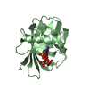
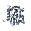
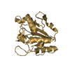
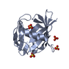
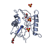
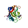
| Title | Crystal structure of high affinity alphaL I domain with ligand mimetic crystal contact | ||||||
 Components Components | Integrin alpha-L | ||||||
 Keywords Keywords |  IMMUNE SYSTEM / Designed disulfide bridge / Rossmann fold / metal mediated protein interface IMMUNE SYSTEM / Designed disulfide bridge / Rossmann fold / metal mediated protein interface | ||||||
| Function / homology |  Function and homology information Function and homology informationmemory T cell extravasation / integrin alphaL-beta2 complex / T cell activation via T cell receptor contact with antigen bound to MHC molecule on antigen presenting cell / ICAM-3 receptor activity / RUNX3 Regulates Immune Response and Cell Migration / integrin complex / cell adhesion mediated by integrin / heterophilic cell-cell adhesion via plasma membrane cell adhesion molecules / leukocyte cell-cell adhesion / receptor clustering ...memory T cell extravasation / integrin alphaL-beta2 complex / T cell activation via T cell receptor contact with antigen bound to MHC molecule on antigen presenting cell / ICAM-3 receptor activity / RUNX3 Regulates Immune Response and Cell Migration / integrin complex / cell adhesion mediated by integrin / heterophilic cell-cell adhesion via plasma membrane cell adhesion molecules / leukocyte cell-cell adhesion / receptor clustering / Integrin cell surface interactions / specific granule membrane / phagocytosis / cell adhesion molecule binding / cell-matrix adhesion / integrin-mediated signaling pathway / Cell surface interactions at the vascular wall / cell-cell adhesion / Immunoregulatory interactions between a Lymphoid and a non-Lymphoid cell / integrin binding / cell adhesion / inflammatory response / external side of plasma membrane / Neutrophil degranulation / cell surface / signal transduction / extracellular exosome / membrane / metal ion binding / plasma membraneSimilarity search - Function | ||||||
| Biological species |  Homo sapiens (human) Homo sapiens (human) | ||||||
| Method | X-RAY DIFFRACTION / SYNCHROTRON / MOLECULAR REPLACEMENT / Resolution: 2 Å | ||||||
 Authors Authors | Shimaoka, M. / Xiao, T. / Liu, J.-H. / Yang, Y. / Dong, Y. / Jun, C.-D. / McCormack, A. / Zhang, R. / Joachimiak, A. / Takagi, J. ...Shimaoka, M. / Xiao, T. / Liu, J.-H. / Yang, Y. / Dong, Y. / Jun, C.-D. / McCormack, A. / Zhang, R. / Joachimiak, A. / Takagi, J. / Wang, J.-H. / Springer, T.A. | ||||||
 Citation Citation | Journal: Cell(Cambridge,Mass.) / Year: 2003 Title: Structures of the aL I domain and its complex with ICAM-1 reveal a shape-shifting pathway for integrin regulation Authors: Shimaoka, M. / Xiao, T. / Liu, J.-H. / Yang, Y. / Dong, Y. / Jun, C.-D. / McCormack, A. / Zhang, R. / Joachimiak, A. / Takagi, J. / Wang, J.-H. / Springer, T.A. | ||||||
| History |
|
- Structure visualization
| Structure viewer | Molecule: MolmilJmol/JSmol |
|---|
- Downloads & links
-Download
| PDBx/mmCIF format | 1mq9.cif.gz | 47.1 KB | Display | PDBx/mmCIF format |
|---|---|---|---|---|
| PDB format | pdb1mq9.ent.gz | 35.9 KB | Display | PDB format |
| PDBx/mmJSON format | 1mq9.json.gz | Tree view | PDBx/mmJSON format | |
| Others |  Other downloads Other downloads |
-Validation report
| Arichive directory | https://data.pdbj.org/pub/pdb/validation_reports/mq/1mq9ftp://data.pdbj.org/pub/pdb/validation_reports/mq/1mq9 | HTTPS FTP |
|---|
-Related structure data
| Related structure data |  1mjnSC 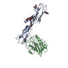 1mq8C  1mqaC C: citing same article ( S: Starting model for refinement |
|---|---|
| Similar structure data |
-Links
 PDBj
PDBj


- Assembly
| Deposited unit | 
| ||||||||||
|---|---|---|---|---|---|---|---|---|---|---|---|
| 1 |
| ||||||||||
| Unit cell |
| ||||||||||
| Components on special symmetry positions |
|
-Components
| #1: Protein | Mass: 20495.502 Da / Num. of mol.: 1 / Fragment: Integrin alphaL I domain / Mutation: K287C, K294C Source method: isolated from a genetically manipulated source Source: (gene. exp.) Homo sapiens (human) / Gene: LFA-1 (AlphaLbeta2) / Plasmid: pET11 / Species (production host): Escherichia coli / Production host:  Escherichia coli BL21(DE3) (bacteria) / Strain (production host): BL21(DE3) / References: UniProt: P20701 Escherichia coli BL21(DE3) (bacteria) / Strain (production host): BL21(DE3) / References: UniProt: P20701 |
|---|---|
| #2: Chemical | ChemComp-MN /   Mass: 54.938 Da / Num. of mol.: 1 / Source method: obtained synthetically / Formula: Mn Mass: 54.938 Da / Num. of mol.: 1 / Source method: obtained synthetically / Formula: Mn |
| #3: Water | ChemComp-HOH / Water Mass: 18.015 Da / Num. of mol.: 90 / Source method: isolated from a natural source / Formula: H2O Mass: 18.015 Da / Num. of mol.: 90 / Source method: isolated from a natural source / Formula: H2O |
-Experimental details
-Experiment
| Experiment | Method: X-RAY DIFFRACTION / Number of used crystals: 1 |
|---|
- Sample preparation
| Crystal | Density Matthews: 2.03 Å3/Da / Density % sol: 38.95 % | |||||||||||||||||||||||||||||||||||
|---|---|---|---|---|---|---|---|---|---|---|---|---|---|---|---|---|---|---|---|---|---|---|---|---|---|---|---|---|---|---|---|---|---|---|---|---|
| Crystal grow | Temperature: 298 K / Method: vapor diffusion, hanging drop / pH: 7 Details: 20% PEG 2000 MME, 25 mM MnCl2, pH 7.0, VAPOR DIFFUSION, HANGING DROP at 298K | |||||||||||||||||||||||||||||||||||
| Crystal grow | *PLUS | |||||||||||||||||||||||||||||||||||
| Components of the solutions | *PLUS
|
-Data collection
| Diffraction | Mean temperature: 100 K |
|---|---|
| Diffraction source | Source: SYNCHROTRON / Site: APS  / Beamline: 19-ID / Wavelength: 1.07 Å / Beamline: 19-ID / Wavelength: 1.07 Å |
| Detector | Type: CUSTOM-MADE / Detector: CCD / Date: Oct 10, 2001 / Details: mirrors |
| Radiation | Monochromator: Ni MIRROR + Ni FILTER / Protocol: SINGLE WAVELENGTH / Monochromatic (M) / Laue (L): M / Scattering type: x-ray |
| Radiation wavelength | Wavelength: 1.07 Å / Relative weight: 1 |
| Reflection | Resolution: 2→50 Å / Num. all: 12800 / Num. obs: 10569 / % possible obs: 82 % / Observed criterion σ(I): -3 / Redundancy: 5.4 % / Biso Wilson estimate: 10.2 Å2 / Rmerge(I) obs: 0.094 / Rsym value: 0.094 / Net I/σ(I): 12.6 |
| Reflection shell | Resolution: 2→2.07 Å / Redundancy: 3.5 % / Rmerge(I) obs: 0.381 / Mean I/σ(I) obs: 3.5 / Num. unique all: 827 / Rsym value: 0.381 / % possible all: 67.5 |
| Reflection | *PLUS Lowest resolution: 50 Å / % possible obs: 82.1 % / Num. measured all: 57584 |
| Reflection shell | *PLUS % possible obs: 67.5 % |
- Processing
| Software |
| |||||||||||||||||||||||||
|---|---|---|---|---|---|---|---|---|---|---|---|---|---|---|---|---|---|---|---|---|---|---|---|---|---|---|
| Refinement | Method to determine structure: MOLECULAR REPLACEMENT Starting model: 1MJN Resolution: 2→27.88 Å / Rfactor Rfree error: 0.008 / Isotropic thermal model: OVERALL / Cross valid method: THROUGHOUT / σ(F): 0 / Stereochemistry target values: Engh & Huber / Details: maximum likelihood refinement target
| |||||||||||||||||||||||||
| Solvent computation | Solvent model: FLAT MODEL / Bsol: 17.8994 Å2 / ksol: 0.315868 e/Å3 | |||||||||||||||||||||||||
| Displacement parameters | Biso mean: 23.8 Å2
| |||||||||||||||||||||||||
| Refine analyze |
| |||||||||||||||||||||||||
| Refinement step | Cycle: LAST / Resolution: 2→27.88 Å
| |||||||||||||||||||||||||
| Refine LS restraints |
| |||||||||||||||||||||||||
| LS refinement shell | Resolution: 2→2.13 Å / Rfactor Rfree error: 0.024 / Total num. of bins used: 6
| |||||||||||||||||||||||||
| Xplor file |
| |||||||||||||||||||||||||
| Refinement | *PLUS Highest resolution: 2 Å / Lowest resolution: 50 Å / % reflection Rfree: 10 % / Rfactor Rwork: 0.213 | |||||||||||||||||||||||||
| Solvent computation | *PLUS | |||||||||||||||||||||||||
| Displacement parameters | *PLUS | |||||||||||||||||||||||||
| Refine LS restraints | *PLUS
|
