 Movie
Movie Controller


+ Open data

- Basic information
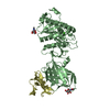
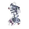
| Entry | Database: PDB / ID: 1mq8 | |||||||||
|---|---|---|---|---|---|---|---|---|---|---|
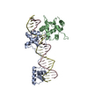
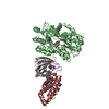
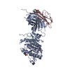
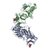
| Title | Crystal structure of alphaL I domain in complex with ICAM-1 | |||||||||
 Components Components |
| |||||||||
 Keywords Keywords |  IMMUNE SYSTEM / Ig superfamily / Rossmann fold / metal mediated protein interface IMMUNE SYSTEM / Ig superfamily / Rossmann fold / metal mediated protein interface | |||||||||
| Function / homology |  Function and homology information Function and homology informationregulation of leukocyte mediated cytotoxicity / T cell extravasation / positive regulation of cellular extravasation / regulation of ruffle assembly / memory T cell extravasation / T cell antigen processing and presentation / integrin alphaL-beta2 complex / T cell activation via T cell receptor contact with antigen bound to MHC molecule on antigen presenting cell / ICAM-3 receptor activity / membrane to membrane docking ...regulation of leukocyte mediated cytotoxicity / T cell extravasation / positive regulation of cellular extravasation / regulation of ruffle assembly / memory T cell extravasation / T cell antigen processing and presentation / integrin alphaL-beta2 complex / T cell activation via T cell receptor contact with antigen bound to MHC molecule on antigen presenting cell / ICAM-3 receptor activity / membrane to membrane docking / adhesion of symbiont to host / RUNX3 Regulates Immune Response and Cell Migration / establishment of endothelial barrier / integrin complex / cell adhesion mediated by integrin / heterophilic cell-cell adhesion via plasma membrane cell adhesion molecules / leukocyte cell-cell adhesion / leukocyte migration / receptor clustering / Interleukin-10 signaling / immunological synapse / Integrin cell surface interactions / specific granule membrane / phagocytosis / negative regulation of endothelial cell apoptotic process / negative regulation of extrinsic apoptotic signaling pathway via death domain receptors / cellular response to leukemia inhibitory factor / cell adhesion molecule binding / cell-matrix adhesion / integrin-mediated signaling pathway / cellular response to glucose stimulus / Cell surface interactions at the vascular wall / cell-cell adhesion / cellular response to amyloid-beta / Immunoregulatory interactions between a Lymphoid and a non-Lymphoid cell / Interferon gamma signaling / transmembrane signaling receptor activity / integrin binding / virus receptor activity / signaling receptor activity / collagen-containing extracellular matrix / Interleukin-4 and Interleukin-13 signaling / receptor-mediated virion attachment to host cell / positive regulation of ERK1 and ERK2 cascade / cell adhesion / inflammatory response / membrane raft / external side of plasma membrane / focal adhesion / Neutrophil degranulation / cell surface / signal transduction / extracellular space / extracellular exosome / membrane / metal ion binding / plasma membraneSimilarity search - Function | |||||||||
| Biological species |  Homo sapiens (human) Homo sapiens (human) | |||||||||
| Method | X-RAY DIFFRACTION / SYNCHROTRON / MOLECULAR REPLACEMENT / Resolution: 3.3 Å | |||||||||
 Authors Authors | Shimaoka, M. / Xiao, T. / Liu, J.-H. / Yang, Y. / Dong, Y. / Jun, C.-D. / McCormack, A. / Zhang, R. / Joachimiak, A. / Takagi, J. ...Shimaoka, M. / Xiao, T. / Liu, J.-H. / Yang, Y. / Dong, Y. / Jun, C.-D. / McCormack, A. / Zhang, R. / Joachimiak, A. / Takagi, J. / Wang, J.-H. / Springer, T.A. | |||||||||
 Citation Citation | Journal: Cell(Cambridge,Mass.) / Year: 2003 Title: Structures of the aL I domain and its complex with ICAM-1 reveal a shape-shifting pathway for integrin regulation Authors: Shimaoka, M. / Xiao, T. / Liu, J.-H. / Yang, Y. / Dong, Y. / Jun, C.-D. / McCormack, A. / Zhang, R. / Joachimiak, A. / Takagi, J. / Wang, J.-H. / Springer, T.A. | |||||||||
| History |
|
- Structure visualization
| Structure viewer | Molecule: MolmilJmol/JSmol |
|---|
- Downloads & links
-Download
| PDBx/mmCIF format | 1mq8.cif.gz | 143.3 KB | Display | PDBx/mmCIF format |
|---|---|---|---|---|
| PDB format | pdb1mq8.ent.gz | 117.4 KB | Display | PDB format |
| PDBx/mmJSON format | 1mq8.json.gz | Tree view | PDBx/mmJSON format | |
| Others |  Other downloads Other downloads |
-Validation report
| Arichive directory | https://data.pdbj.org/pub/pdb/validation_reports/mq/1mq8ftp://data.pdbj.org/pub/pdb/validation_reports/mq/1mq8 | HTTPS FTP |
|---|
-Related structure data
| Related structure data |  1mjnC  1mq9C  1mqaC  1ic1S C: citing same article ( S: Starting model for refinement |
|---|---|
| Similar structure data |


-Links
 PDBj
PDBj


- Assembly
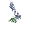
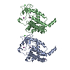
| Deposited unit | 
| ||||||||||
|---|---|---|---|---|---|---|---|---|---|---|---|
| 1 | 
| ||||||||||
| 2 | 
| ||||||||||
| Unit cell |
| ||||||||||
| Details | chain A and B form a biological complex, so do chain C and D. Chain A and C also form a biological dimer. |
-Components
-Protein , 2 types, 4 molecules ACBD
| #1: Protein | / ICAM-1 / CD54 antigen / Major group rhinovirus receptor Mass: 31623.771 Da / Num. of mol.: 2 / Fragment: domains 1 and 2 Source method: isolated from a genetically manipulated source Source: (gene. exp.) Homo sapiens (human) / Gene: ICAM-1 / Plasmid: pMT/BiP/V5-His / Production host: Schneider S2 insect cells / Strain (production host): Schneider S2 insect cells / References: UniProt: P05362#2: Protein | Mass: 20191.178 Da / Num. of mol.: 2 / Fragment: Integrin alphaL I domain / Mutation: L186C, F324C Source method: isolated from a genetically manipulated source Source: (gene. exp.) Homo sapiens (human) / Gene: LFA-1 (AlphaLbeta2) / Plasmid: pET11 / Species (production host): Escherichia coli / Production host:  Escherichia coli BL21(DE3) (bacteria) / Strain (production host): BL21(DE3) / References: UniProt: P20701 Escherichia coli BL21(DE3) (bacteria) / Strain (production host): BL21(DE3) / References: UniProt: P20701 |
|---|
-Sugars , 2 types, 8 molecules 
| #3: Polysaccharide | 2-acetamido-2-deoxy-beta-D-glucopyranose-(1-4)-2-acetamido-2-deoxy-beta-D-glucopyranose / Mass: 424.401 Da / Num. of mol.: 4Source method: isolated from a genetically manipulated source #4: Sugar | ChemComp-NAG / N-Acetylglucosamine Type: D-saccharide, beta linking / Mass: 221.208 Da / Num. of mol.: 4 Type: D-saccharide, beta linking / Mass: 221.208 Da / Num. of mol.: 4Source method: isolated from a genetically manipulated source Formula: C8H15NO6 |
|---|
-Non-polymers , 2 types, 6 molecules 

| #5: Chemical |  Mass: 24.305 Da / Num. of mol.: 2 / Source method: obtained synthetically / Formula: Mg Mass: 24.305 Da / Num. of mol.: 2 / Source method: obtained synthetically / Formula: Mg#6: Water | ChemComp-HOH / | WaterMass: 18.015 Da / Num. of mol.: 4 / Source method: isolated from a natural source / Formula: H2O |
|---|
-Experimental details
-Experiment
| Experiment | Method: X-RAY DIFFRACTION / Number of used crystals: 1 |
|---|
- Sample preparation
| Crystal | Density Matthews: 2.78 Å3/Da / Density % sol: 55.3 % | ||||||||||||||||||||||||
|---|---|---|---|---|---|---|---|---|---|---|---|---|---|---|---|---|---|---|---|---|---|---|---|---|---|
| Crystal grow | Temperature: 298 K / Method: vapor diffusion, hanging drop / pH: 4.6 Details: 25% PEG 4000, 0.1 M sodium acetate, pH 4.6, VAPOR DIFFUSION, HANGING DROP at 298K | ||||||||||||||||||||||||
| Crystal grow | *PLUS | ||||||||||||||||||||||||
| Components of the solutions | *PLUS
|
-Data collection
| Diffraction | Mean temperature: 100 K |
|---|---|
| Diffraction source | Source: SYNCHROTRON / Site: APS  / Beamline: 19-ID / Wavelength: 0.91 Å / Beamline: 19-ID / Wavelength: 0.91 Å |
| Detector | Type: CUSTOM-MADE / Detector: CCD / Date: Jan 1, 2001 / Details: mirrors |
| Radiation | Monochromator: Ni MIRROR + Ni FILTER / Protocol: SINGLE WAVELENGTH / Monochromatic (M) / Laue (L): M / Scattering type: x-ray |
| Radiation wavelength | Wavelength: 0.91 Å / Relative weight: 1 |
| Reflection | Resolution: 3.3→50 Å / Num. all: 25280 / Num. obs: 23330 / % possible obs: 92.3 % / Observed criterion σ(I): -3 / Redundancy: 1.9 % / Biso Wilson estimate: 46.4 Å2 / Rmerge(I) obs: 0.094 / Rsym value: 0.094 / Net I/σ(I): 7.7 |
| Reflection shell | Resolution: 3.3→3.36 Å / Redundancy: 1 % / Rmerge(I) obs: 0.412 / Mean I/σ(I) obs: 1.5 / Num. unique all: 469 / Rsym value: 0.412 / % possible all: 70.7 |
| Reflection | *PLUS Highest resolution: 3.3 Å / Lowest resolution: 50 Å / Num. obs: 12338 / Num. measured all: 23330 |
| Reflection shell | *PLUS % possible obs: 70.7 % |
- Processing
| Software |
| |||||||||||||||||||||||||
|---|---|---|---|---|---|---|---|---|---|---|---|---|---|---|---|---|---|---|---|---|---|---|---|---|---|---|
| Refinement | Method to determine structure: MOLECULAR REPLACEMENT Starting model: 1IC1 Resolution: 3.3→50 Å / Rfactor Rfree error: 0.013 / Isotropic thermal model: Isotropic / Cross valid method: THROUGHOUT / σ(F): 0 / σ(I): 0 / Stereochemistry target values: Engh & Huber / Details: maximum likelihood refinement target
| |||||||||||||||||||||||||
| Displacement parameters | Biso mean: 67 Å2
| |||||||||||||||||||||||||
| Refine analyze |
| |||||||||||||||||||||||||
| Refinement step | Cycle: LAST / Resolution: 3.3→50 Å
| |||||||||||||||||||||||||
| Refine LS restraints |
| |||||||||||||||||||||||||
| Refinement | *PLUS Highest resolution: 3.3 Å / % reflection Rfree: 5 % | |||||||||||||||||||||||||
| Solvent computation | *PLUS | |||||||||||||||||||||||||
| Displacement parameters | *PLUS | |||||||||||||||||||||||||
| Refine LS restraints | *PLUS
|
