 Movie
Movie Controller

[English] 日本語
 Yorodumi
- PDB-1mcp: PHOSPHOCHOLINE BINDING IMMUNOGLOBULIN FAB MC/PC603. AN X-RAY DIFF... -
+ Open data

- Basic information
| Entry | Database: PDB / ID: 1mcp | |||||||||
|---|---|---|---|---|---|---|---|---|---|---|
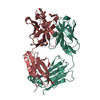
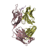
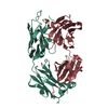
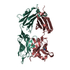
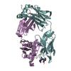
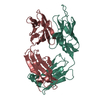
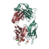
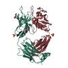
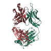
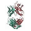
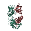
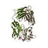
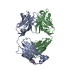
| Title | PHOSPHOCHOLINE BINDING IMMUNOGLOBULIN FAB MC/PC603. AN X-RAY DIFFRACTION STUDY AT 2.7 ANGSTROMS | |||||||||
 Components Components |
| |||||||||
 Keywords Keywords |  IMMUNOGLOBULIN IMMUNOGLOBULIN | |||||||||
| Function / homology |  Function and homology informationimmunoglobulin complex / immunoglobulin mediated immune response / antigen binding Function and homology informationimmunoglobulin complex / immunoglobulin mediated immune response / antigen bindingSimilarity search - Function | |||||||||
| Biological species |  Mus musculus (house mouse) Mus musculus (house mouse) | |||||||||
| Method | X-RAY DIFFRACTION / Resolution: 2.7 Å | |||||||||
 Authors Authors | Satow, Y. / Cohen, G.H. / Padlan, E.A. / Davies, D.R. | |||||||||
 Citation Citation | Journal: J.Mol.Biol. / Year: 1986 Title: Phosphocholine binding immunoglobulin Fab McPC603. An X-ray diffraction study at 2.7 A. Authors: Satow, Y. / Cohen, G.H. / Padlan, E.A. / Davies, D.R. #1: Journal: Mol.Immunol. / Year: 1981Title: Kappa Chain Structure from a Crystallized Murine Fab(Prime). Role of Joining Segment in Hapten Binding Authors: Rudikoff, S. / Satow, Y. / Padlan, E. / Davies, D. / Potter, M. #2: Journal: Contemp.Top.Mol.Immunol. / Year: 1975Title: Immunoglobulin Structures at High Resolution Authors: Davies, D.R. / Padlan, E.A. / Segal, D.M. #3: Journal: The Immune System. Genes,Receptors,Signals. Proceedings of the 1974 I.C.N.-U.C.L.A. Symposium on Molecular BiologyYear: 1974 Title: The Three-Dimensional Structure of the Antigen Binding Site of Mc/Pc 603 Protein Authors: Padlan, E.A. / Segal, D.M. / Cohen, G.H. / Davies, D.R. / Rudikoff, S. / Potter, M. #4: Journal: Proc.Natl.Acad.Sci.USA / Year: 1974Title: The Three-Dimensional Structure of a Phosphorylcholine-Binding Mouse Immunoglobulin Fab and the Nature of the Antigen Binding Site Authors: Segal, D.M. / Padlan, E.A. / Cohen, G.H. / Rudikoff, S. / Potter, M. / Davies, D.R. #5: Journal: Nature New Biol. / Year: 1973Title: Structure at 4.5 Angstroms Resolution of a Phosphorylcholine-Binding Fab Authors: Padlan, E.A. / Segal, D.M. / Spande, T.F. / Davies, D.R. / Rudikoff, S. / Potter, M. #6: Journal: Proc.Natl.Acad.Sci.USA / Year: 1972Title: Crystals of Phosphorylcholine-Binding Fab-Fragments from Mouse Myeloma Proteins. Preparation and X-Ray Analysis Authors: Rudikoff, S. / Potter, M. / Segal, D.M. / Padlan, E.A. / Davies, D.R. | |||||||||
| History |
|
- Structure visualization
| Structure viewer | Molecule: MolmilJmol/JSmol |
|---|
- Downloads & links
-Download
| PDBx/mmCIF format | 1mcp.cif.gz | 98.3 KB | Display | PDBx/mmCIF format |
|---|---|---|---|---|
| PDB format | pdb1mcp.ent.gz | 77.6 KB | Display | PDB format |
| PDBx/mmJSON format | 1mcp.json.gz | Tree view | PDBx/mmJSON format | |
| Others |  Other downloads Other downloads |
-Validation report
| Arichive directory | https://data.pdbj.org/pub/pdb/validation_reports/mc/1mcpftp://data.pdbj.org/pub/pdb/validation_reports/mc/1mcp | HTTPS FTP |
|---|
-Related structure data
| Similar structure data |
|---|


-Links
 PDBj
PDBj

- Assembly
| Deposited unit | 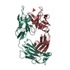
| ||||||||
|---|---|---|---|---|---|---|---|---|---|
| 1 |
| ||||||||
| Unit cell |
| ||||||||
| Atom site foot note | 1: FOR RESIDUES 28-37 AND 162-163 OF THE LIGHT CHAIN AND RESIDUE 202 OF THE HEAVY CHAIN, THE DENSITY WAS POOR. THESE RESIDUES WERE BUILT MAINLY BY STEREOCHEMISTRY. 2: RESIDUES PRO L 8, PRO L 101, PRO L 147, PRO H 143, PRO H 155 ARE CIS PROLINES. |
-Components
| #1: Antibody | Mass: 24113.584 Da / Num. of mol.: 1 Source method: isolated from a genetically manipulated source Source: (gene. exp.) Mus musculus (house mouse) / References: GenBank: 208622 |
|---|---|
| #2: Antibody | Mass: 24319.266 Da / Num. of mol.: 1 Source method: isolated from a genetically manipulated source Source: (gene. exp.) Mus musculus (house mouse) / References: UniProt: P01789 |
| #3: Chemical | ChemComp-SO4 / Sulfate  Mass: 96.063 Da / Num. of mol.: 1 / Source method: obtained synthetically / Formula: SO4 Mass: 96.063 Da / Num. of mol.: 1 / Source method: obtained synthetically / Formula: SO4 |
| #4: Water | ChemComp-HOH / Water Mass: 18.015 Da / Num. of mol.: 138 / Source method: isolated from a natural source / Formula: H2O Mass: 18.015 Da / Num. of mol.: 138 / Source method: isolated from a natural source / Formula: H2O |
-Experimental details
-Experiment
| Experiment | Method: X-RAY DIFFRACTION |
|---|
- Sample preparation
| Crystal | Density Matthews: 4.78 Å3/Da / Density % sol: 74.25 % | |||||||||||||||
|---|---|---|---|---|---|---|---|---|---|---|---|---|---|---|---|---|
| Crystal grow | *PLUS pH: 7 / Method: vapor diffusion | |||||||||||||||
| Components of the solutions | *PLUS
|
-Data collection
| Radiation | Scattering type: x-ray |
|---|---|
| Radiation wavelength | Relative weight: 1 |
| Reflection | *PLUS Highest resolution: 2.7 Å / Lowest resolution: 10 Å / Num. obs: 24235 / Num. measured all: 147706 |
- Processing
| Refinement | Rfactor Rwork: 0.225 / Highest resolution: 2.7 Å | ||||||||||||||||||||||||||||||||||||||||||||||||||||||||||||
|---|---|---|---|---|---|---|---|---|---|---|---|---|---|---|---|---|---|---|---|---|---|---|---|---|---|---|---|---|---|---|---|---|---|---|---|---|---|---|---|---|---|---|---|---|---|---|---|---|---|---|---|---|---|---|---|---|---|---|---|---|---|
| Refinement step | Cycle: LAST / Highest resolution: 2.7 Å
| ||||||||||||||||||||||||||||||||||||||||||||||||||||||||||||
| Refine LS restraints |
| ||||||||||||||||||||||||||||||||||||||||||||||||||||||||||||
| Refinement | *PLUS Highest resolution: 2.7 Å / Lowest resolution: 8 Å / Num. reflection obs: 23737 / Rfactor obs: 0.225 | ||||||||||||||||||||||||||||||||||||||||||||||||||||||||||||
| Solvent computation | *PLUS | ||||||||||||||||||||||||||||||||||||||||||||||||||||||||||||
| Displacement parameters | *PLUS | ||||||||||||||||||||||||||||||||||||||||||||||||||||||||||||
| Refine LS restraints | *PLUS
|
