 Movie
Movie Controller


+ Open data

- Basic information
| Entry | Database: PDB / ID: 1m4s | ||||||
|---|---|---|---|---|---|---|---|
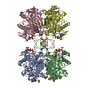
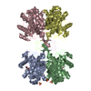
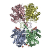
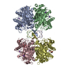
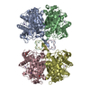
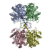
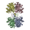
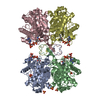
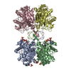
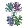
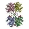
| Title | Biosynthetic thiolase, Cys89 acetylated, unliganded form | ||||||
 Components Components | Acetyl-CoA acetyltransferase | ||||||
 Keywords Keywords |  TRANSFERASE / thiolase fold / acetylated intermediate TRANSFERASE / thiolase fold / acetylated intermediate | ||||||
| Function / homology |  Function and homology information Function and homology informationpoly-hydroxybutyrate biosynthetic process / acetyl-CoA C-acetyltransferase / acetyl-CoA C-acetyltransferase activity / cytoplasmSimilarity search - Function | ||||||
| Biological species |  Zoogloea ramigera (bacteria) Zoogloea ramigera (bacteria) | ||||||
| Method | X-RAY DIFFRACTION / SYNCHROTRON / MOLECULAR REPLACEMENT / Resolution: 1.87 Å | ||||||
 Authors Authors | Kursula, P. / Ojala, J. / Lambeir, A.-M. / Wierenga, R.K. | ||||||
 Citation Citation | Journal: Biochemistry / Year: 2002 Title: The catalytic cycle of biosynthetic thiolase: A conformational journey of an acetyl group through four binding modes and two oxyanion holes Authors: Kursula, P. / Ojala, J. / Lambeir, A.-M. / Wierenga, R.K. | ||||||
| History |
|
- Structure visualization
| Structure viewer | Molecule: MolmilJmol/JSmol |
|---|
- Downloads & links
-Download
| PDBx/mmCIF format | 1m4s.cif.gz | 306.8 KB | Display | PDBx/mmCIF format |
|---|---|---|---|---|
| PDB format | pdb1m4s.ent.gz | 257.6 KB | Display | PDB format |
| PDBx/mmJSON format | 1m4s.json.gz | Tree view | PDBx/mmJSON format | |
| Others |  Other downloads Other downloads |
-Validation report
| Arichive directory | https://data.pdbj.org/pub/pdb/validation_reports/m4/1m4sftp://data.pdbj.org/pub/pdb/validation_reports/m4/1m4s | HTTPS FTP |
|---|
-Related structure data
| Related structure data |  1m1oC 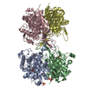 1m1tC  1m3kC  1m3zC  1m4tC  1dluS S: Starting model for refinement C: citing same article ( |
|---|---|
| Similar structure data |


-Links
 PDBj
PDBj

- Assembly
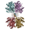
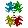
| Deposited unit | 
| ||||||||||
|---|---|---|---|---|---|---|---|---|---|---|---|
| 1 |
| ||||||||||
| Unit cell |
|
-Components
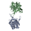
| #1: Protein | Mass: 40583.246 Da / Num. of mol.: 4 / Mutation: C89 acetylated Source method: isolated from a genetically manipulated source Source: (gene. exp.) Zoogloea ramigera (bacteria) / Production host: Escherichia coli (E. coli) / References: UniProt: P07097, acetyl-CoA C-acetyltransferase#2: Chemical | ChemComp-SO4 / Sulfate  Mass: 96.063 Da / Num. of mol.: 4 / Source method: obtained synthetically / Formula: SO4 Mass: 96.063 Da / Num. of mol.: 4 / Source method: obtained synthetically / Formula: SO4#3: Chemical | ChemComp-GOL / Glycerol  Mass: 92.094 Da / Num. of mol.: 4 / Source method: obtained synthetically / Formula: C3H8O3 Mass: 92.094 Da / Num. of mol.: 4 / Source method: obtained synthetically / Formula: C3H8O3#4: Water | ChemComp-HOH / | Water Mass: 18.015 Da / Num. of mol.: 1094 / Source method: isolated from a natural source / Formula: H2O Mass: 18.015 Da / Num. of mol.: 1094 / Source method: isolated from a natural source / Formula: H2O |
|---|
-Experimental details
-Experiment
| Experiment | Method: X-RAY DIFFRACTION / Number of used crystals: 1 |
|---|
- Sample preparation
| Crystal | Density Matthews: 3.06 Å3/Da / Density % sol: 59.76 % | ||||||||||||||||||||||||||||||||||||||||||||||||||||||||
|---|---|---|---|---|---|---|---|---|---|---|---|---|---|---|---|---|---|---|---|---|---|---|---|---|---|---|---|---|---|---|---|---|---|---|---|---|---|---|---|---|---|---|---|---|---|---|---|---|---|---|---|---|---|---|---|---|---|
| Crystal grow | Temperature: 293 K / Method: vapor diffusion, sitting drop / pH: 5 Details: lithium sulphate, ammonium sulphate, pH 5.0, VAPOR DIFFUSION, SITTING DROP at 293K | ||||||||||||||||||||||||||||||||||||||||||||||||||||||||
| Crystal grow | *PLUS Method: vapor diffusion, hanging drop | ||||||||||||||||||||||||||||||||||||||||||||||||||||||||
| Components of the solutions | *PLUS
|
-Data collection
| Diffraction | Mean temperature: 100 K |
|---|---|
| Diffraction source | Source: SYNCHROTRON / Site: MAX II  / Beamline: I711 / Wavelength: 1.09785 Å / Beamline: I711 / Wavelength: 1.09785 Å |
| Detector | Type: MARRESEARCH / Detector: CCD / Date: Sep 22, 2001 |
| Radiation | Protocol: SINGLE WAVELENGTH / Monochromatic (M) / Laue (L): M / Scattering type: x-ray |
| Radiation wavelength | Wavelength: 1.09785 Å / Relative weight: 1 |
| Reflection | Resolution: 1.87→50 Å / Num. all: 158418 / Num. obs: 158418 / % possible obs: 97.9 % / Observed criterion σ(I): -3 / Redundancy: 3.3 % / Biso Wilson estimate: 24 Å2 / Rsym value: 0.092 / Net I/σ(I): 9.5 |
| Reflection shell | Resolution: 1.87→1.95 Å / Redundancy: 2.6 % / Mean I/σ(I) obs: 3.2 / Num. unique all: 17066 / Rsym value: 0.354 / % possible all: 90.1 |
| Reflection | *PLUS Rmerge(I) obs: 0.092 |
| Reflection shell | *PLUS % possible obs: 90.1 % / Rmerge(I) obs: 0.354 |
- Processing
| Software |
| ||||||||||||||||||||||||||||||||||||||||||||||||||||||||||||||||||||||||||||||||||||||||||||||||||||||||||||||||||||||||||||||||||
|---|---|---|---|---|---|---|---|---|---|---|---|---|---|---|---|---|---|---|---|---|---|---|---|---|---|---|---|---|---|---|---|---|---|---|---|---|---|---|---|---|---|---|---|---|---|---|---|---|---|---|---|---|---|---|---|---|---|---|---|---|---|---|---|---|---|---|---|---|---|---|---|---|---|---|---|---|---|---|---|---|---|---|---|---|---|---|---|---|---|---|---|---|---|---|---|---|---|---|---|---|---|---|---|---|---|---|---|---|---|---|---|---|---|---|---|---|---|---|---|---|---|---|---|---|---|---|---|---|---|---|---|
| Refinement | Method to determine structure: MOLECULAR REPLACEMENT Starting model: 1DLU Resolution: 1.87→20 Å / Cor.coef. Fo:Fc: 0.923 / Cor.coef. Fo:Fc free: 0.891 / TLS residual ADP flag: LIKELY RESIDUAL Isotropic thermal model: ISOTROPIC REFINEMENT, TLS PARAMETERISATION Cross valid method: THROUGHOUT / σ(F): 0 / σ(I): -3 / Stereochemistry target values: Engh & Huber Details: HYDROGENS HAVE BEEN ADDED IN THE RIDING POSITIONS DURING REFINEMENT
| ||||||||||||||||||||||||||||||||||||||||||||||||||||||||||||||||||||||||||||||||||||||||||||||||||||||||||||||||||||||||||||||||||
| Solvent computation | Shrinkage radii: 0.8 Å / Solvent model: BABINET MODEL WITH MASK | ||||||||||||||||||||||||||||||||||||||||||||||||||||||||||||||||||||||||||||||||||||||||||||||||||||||||||||||||||||||||||||||||||
| Displacement parameters | Biso mean: 12.727 Å2
| ||||||||||||||||||||||||||||||||||||||||||||||||||||||||||||||||||||||||||||||||||||||||||||||||||||||||||||||||||||||||||||||||||
| Refinement step | Cycle: LAST / Resolution: 1.87→20 Å
| ||||||||||||||||||||||||||||||||||||||||||||||||||||||||||||||||||||||||||||||||||||||||||||||||||||||||||||||||||||||||||||||||||
| Refine LS restraints |
| ||||||||||||||||||||||||||||||||||||||||||||||||||||||||||||||||||||||||||||||||||||||||||||||||||||||||||||||||||||||||||||||||||
| LS refinement shell | Resolution: 1.87→1.918 Å / Total num. of bins used: 20 /
| ||||||||||||||||||||||||||||||||||||||||||||||||||||||||||||||||||||||||||||||||||||||||||||||||||||||||||||||||||||||||||||||||||
| Refinement TLS params. | Method: refined / Refine-ID: X-RAY DIFFRACTION
| ||||||||||||||||||||||||||||||||||||||||||||||||||||||||||||||||||||||||||||||||||||||||||||||||||||||||||||||||||||||||||||||||||
| Refinement TLS group |
| ||||||||||||||||||||||||||||||||||||||||||||||||||||||||||||||||||||||||||||||||||||||||||||||||||||||||||||||||||||||||||||||||||
| Refinement | *PLUS Lowest resolution: 20 Å / Num. reflection obs: 151113 / Rfactor Rfree: 0.247 / Rfactor Rwork: 0.206 | ||||||||||||||||||||||||||||||||||||||||||||||||||||||||||||||||||||||||||||||||||||||||||||||||||||||||||||||||||||||||||||||||||
| Solvent computation | *PLUS | ||||||||||||||||||||||||||||||||||||||||||||||||||||||||||||||||||||||||||||||||||||||||||||||||||||||||||||||||||||||||||||||||||
| Displacement parameters | *PLUS | ||||||||||||||||||||||||||||||||||||||||||||||||||||||||||||||||||||||||||||||||||||||||||||||||||||||||||||||||||||||||||||||||||
| Refine LS restraints | *PLUS
|
