 Movie
Movie Controller

[English] 日本語
 Yorodumi
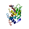
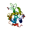
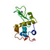
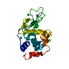
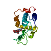
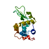
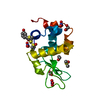
- PDB-1lzs: STRUCTURAL CHANGES OF THE ACTIVE SITE CLEFT AND DIFFERENT SACCHAR... -
+ Open data

- Basic information
| Entry | Database: PDB / ID: 1lzs | |||||||||
|---|---|---|---|---|---|---|---|---|---|---|
| Title | STRUCTURAL CHANGES OF THE ACTIVE SITE CLEFT AND DIFFERENT SACCHARIDE BINDING MODES IN HUMAN LYSOZYME CO-CRYSTALLIZED WITH HEXA-N-ACETYL-CHITOHEXAOSE AT PH 4.0 | |||||||||
 Components Components | HUMAN LYSOZYME | |||||||||
 Keywords Keywords | HYDROLASE (O-GLYCOSYL) | |||||||||
| Function / homology |  Function and homology informationcytolysis / antimicrobial humoral response / retina homeostasis / Antimicrobial peptides / metabolic process / specific granule lumen / azurophil granule lumen / tertiary granule lumen / lysozyme / lysozyme activity ...cytolysis / antimicrobial humoral response / retina homeostasis / Antimicrobial peptides / metabolic process / specific granule lumen / azurophil granule lumen / tertiary granule lumen / lysozyme / lysozyme activity / killing of cells of another organism / defense response to Gram-negative bacterium / defense response to Gram-positive bacterium / defense response to bacterium / inflammatory response / Amyloid fiber formation / Neutrophil degranulation / extracellular space / extracellular exosome / extracellular region / identical protein binding Function and homology informationcytolysis / antimicrobial humoral response / retina homeostasis / Antimicrobial peptides / metabolic process / specific granule lumen / azurophil granule lumen / tertiary granule lumen / lysozyme / lysozyme activity ...cytolysis / antimicrobial humoral response / retina homeostasis / Antimicrobial peptides / metabolic process / specific granule lumen / azurophil granule lumen / tertiary granule lumen / lysozyme / lysozyme activity / killing of cells of another organism / defense response to Gram-negative bacterium / defense response to Gram-positive bacterium / defense response to bacterium / inflammatory response / Amyloid fiber formation / Neutrophil degranulation / extracellular space / extracellular exosome / extracellular region / identical protein bindingSimilarity search - Function | |||||||||
| Biological species |  Homo sapiens (human) Homo sapiens (human) | |||||||||
| Method | X-RAY DIFFRACTION / Resolution: 1.6 Å | |||||||||
 Authors Authors | Matsushima, M. / Song, H. | |||||||||
 Citation Citation | Journal: J.Mol.Biol. / Year: 1994 Title: Structural changes of active site cleft and different saccharide binding modes in human lysozyme co-crystallized with hexa-N-acetyl-chitohexaose at pH 4.0. Authors: Song, H. / Inaka, K. / Maenaka, K. / Matsushima, M. | |||||||||
| History |
|
- Structure visualization
| Structure viewer | Molecule: MolmilJmol/JSmol |
|---|
- Downloads & links
-Download
| PDBx/mmCIF format | 1lzs.cif.gz | 69.5 KB | Display | PDBx/mmCIF format |
|---|---|---|---|---|
| PDB format | pdb1lzs.ent.gz | 55.1 KB | Display | PDB format |
| PDBx/mmJSON format | 1lzs.json.gz | Tree view | PDBx/mmJSON format | |
| Others |  Other downloads Other downloads |
-Validation report
| Arichive directory | https://data.pdbj.org/pub/pdb/validation_reports/lz/1lzsftp://data.pdbj.org/pub/pdb/validation_reports/lz/1lzs | HTTPS FTP |
|---|
-Related structure data


-Links
 PDBj
PDBj

- Assembly
| Deposited unit | 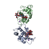
| ||||||||
|---|---|---|---|---|---|---|---|---|---|
| 1 | 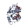
| ||||||||
| 2 | 
| ||||||||
| Unit cell |
|
-Components
| #1: Protein | Mass: 14720.693 Da / Num. of mol.: 2 Source method: isolated from a genetically manipulated source Source: (gene. exp.) Homo sapiens (human) / References: UniProt: P00695, UniProt: P61626*PLUS, lysozyme#2: Polysaccharide | / Mass: 830.786 Da / Num. of mol.: 2Source method: isolated from a genetically manipulated source #3: Polysaccharide | 2-acetamido-2-deoxy-beta-D-glucopyranose-(1-4)-2-acetamido-2-deoxy-beta-D-glucopyranose | / Mass: 424.401 Da / Num. of mol.: 1Source method: isolated from a genetically manipulated source #4: Chemical |   Mass: 22.990 Da / Num. of mol.: 2 / Source method: obtained synthetically / Formula: Na Mass: 22.990 Da / Num. of mol.: 2 / Source method: obtained synthetically / Formula: Na#5: Water | ChemComp-HOH / | Water Mass: 18.015 Da / Num. of mol.: 271 / Source method: isolated from a natural source / Formula: H2O Mass: 18.015 Da / Num. of mol.: 271 / Source method: isolated from a natural source / Formula: H2OCompound details | ONE OF THE LYSOZYME MOLECULES BINDS THE CLEAVED SACCHARIDE MOLECULES, (NAG)4 IN THE BINDING ...ONE OF THE LYSOZYME MOLECULES BINDS THE CLEAVED SACCHARIDE | |
|---|
-Experimental details
-Experiment
| Experiment | Method: X-RAY DIFFRACTION |
|---|
- Sample preparation
| Crystal | Density Matthews: 2.11 Å3/Da / Density % sol: 41.57 % | ||||||||||||||||||||||||||||||||||||||||||
|---|---|---|---|---|---|---|---|---|---|---|---|---|---|---|---|---|---|---|---|---|---|---|---|---|---|---|---|---|---|---|---|---|---|---|---|---|---|---|---|---|---|---|---|
| Crystal grow | *PLUS Temperature: 4 ℃ / pH: 4 / Method: vapor diffusion | ||||||||||||||||||||||||||||||||||||||||||
| Components of the solutions | *PLUS
|
-Data collection
| Radiation | Scattering type: x-ray |
|---|---|
| Radiation wavelength | Relative weight: 1 |
| Reflection | *PLUS Highest resolution: 1.6 Å / Lowest resolution: 9999 Å / Num. obs: 25249 / % possible obs: 74.5 % / Observed criterion σ(I): 2 / Num. measured all: 63078 / Rmerge(I) obs: 0.053 |
| Reflection shell | *PLUS Highest resolution: 1.6 Å / Lowest resolution: 1.63 Å / % possible obs: 45.5 % |
- Processing
| Software | Name: PROLSQ / Classification: refinement | ||||||||||||||||||||||||||||||||||||||||||||||||||||||||||||||||||||||||||||||||||||
|---|---|---|---|---|---|---|---|---|---|---|---|---|---|---|---|---|---|---|---|---|---|---|---|---|---|---|---|---|---|---|---|---|---|---|---|---|---|---|---|---|---|---|---|---|---|---|---|---|---|---|---|---|---|---|---|---|---|---|---|---|---|---|---|---|---|---|---|---|---|---|---|---|---|---|---|---|---|---|---|---|---|---|---|---|---|
| Refinement | Resolution: 1.6→6 Å /
| ||||||||||||||||||||||||||||||||||||||||||||||||||||||||||||||||||||||||||||||||||||
| Refinement step | Cycle: LAST / Resolution: 1.6→6 Å
| ||||||||||||||||||||||||||||||||||||||||||||||||||||||||||||||||||||||||||||||||||||
| Refine LS restraints |
| ||||||||||||||||||||||||||||||||||||||||||||||||||||||||||||||||||||||||||||||||||||
| LS refinement shell | *PLUS Highest resolution: 1.6 Å / Lowest resolution: 1.7 Å / Rfactor all: 0.225 |
