 Movie
Movie Controller


+ Open data

- Basic information
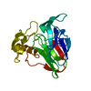
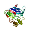
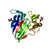
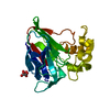
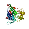
| Entry | Database: PDB / ID: 1ly0 | ||||||
|---|---|---|---|---|---|---|---|
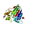
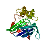
| Title | Structure of thaumatin crystallized in the presence of glycerol | ||||||
 Components Components | Thaumatin I | ||||||
 Keywords Keywords | PLANT PROTEIN / TASTE-MODIFYING PROTEIN / SWEET PROTEIN | ||||||
| Function / homology |  Function and homology information Function and homology information | ||||||
| Biological species |  Thaumatococcus daniellii (katemfe) Thaumatococcus daniellii (katemfe) | ||||||
| Method | X-RAY DIFFRACTION / SYNCHROTRON / MOLECULAR REPLACEMENT / Resolution: 1.36 Å | ||||||
 Authors Authors | Charron, C. / Kadri, A. / Robert, M.C. / Giege, R. / Lorber, B. | ||||||
 Citation Citation | Journal: Acta Crystallogr.,Sect.D / Year: 2002 Title: Crystallization in the presence of glycerol displaces water molecules in the structure of thaumatin. Authors: Charron, C. / Kadri, A. / Robert, M.C. / Giege, R. / Lorber, B. | ||||||
| History |
|
- Structure visualization
| Structure viewer | Molecule: MolmilJmol/JSmol |
|---|
- Downloads & links
-Download
| PDBx/mmCIF format | 1ly0.cif.gz | 53.7 KB | Display | PDBx/mmCIF format |
|---|---|---|---|---|
| PDB format | pdb1ly0.ent.gz | 41.9 KB | Display | PDB format |
| PDBx/mmJSON format | 1ly0.json.gz | Tree view | PDBx/mmJSON format | |
| Others |  Other downloads Other downloads |
-Validation report
| Arichive directory | https://data.pdbj.org/pub/pdb/validation_reports/ly/1ly0ftp://data.pdbj.org/pub/pdb/validation_reports/ly/1ly0 | HTTPS FTP |
|---|
-Related structure data
| Related structure data | 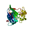 1lxzC 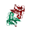 1thwS C: citing same article ( S: Starting model for refinement |
|---|---|
| Similar structure data |


-Links
 PDBj
PDBj

- Assembly
| Deposited unit | 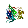
| ||||||||
|---|---|---|---|---|---|---|---|---|---|
| 1 |
| ||||||||
| Unit cell |
| ||||||||
| Details | The biological unit is a monomer |
-Components
| #1: Protein | Mass: 22227.059 Da / Num. of mol.: 1 / Source method: isolated from a natural source / Source: (natural) Thaumatococcus daniellii (katemfe) / References: UniProt: P02883 |
|---|---|
| #2: Chemical | ChemComp-TLA / Tartaric acid  Mass: 150.087 Da / Num. of mol.: 1 / Source method: obtained synthetically / Formula: C4H6O6 Mass: 150.087 Da / Num. of mol.: 1 / Source method: obtained synthetically / Formula: C4H6O6 |
| #3: Water | ChemComp-HOH / Water Mass: 18.015 Da / Num. of mol.: 325 / Source method: isolated from a natural source / Formula: H2O Mass: 18.015 Da / Num. of mol.: 325 / Source method: isolated from a natural source / Formula: H2O |
-Experimental details
-Experiment
| Experiment | Method: X-RAY DIFFRACTION / Number of used crystals: 1 |
|---|
- Sample preparation
| Crystal | Density Matthews: 2.82 Å3/Da / Density % sol: 56.42 % |
|---|---|
| Crystal grow | Temperature: 293 K / Method: vapor diffusion, hanging drop / pH: 6.5 Details: tartrate, pH 6.5, VAPOR DIFFUSION, HANGING DROP, temperature 293K |
-Data collection
| Diffraction | Mean temperature: 100 K |
|---|---|
| Diffraction source | Source: SYNCHROTRON / Site: LURE  / Beamline: DW32 / Wavelength: 0.9474 Å / Beamline: DW32 / Wavelength: 0.9474 Å |
| Detector | Type: MARRESEARCH / Detector: IMAGE PLATE / Date: Dec 17, 2000 |
| Radiation | Protocol: SINGLE WAVELENGTH / Monochromatic (M) / Laue (L): M / Scattering type: x-ray |
| Radiation wavelength | Wavelength: 0.9474 Å / Relative weight: 1 |
| Reflection | Resolution: 1.36→30 Å / Num. all: 55721 / Num. obs: 55482 / % possible obs: 99.9 % / Observed criterion σ(I): -3 / Redundancy: 7.3 % / Rsym value: 0.049 |
| Reflection shell | Resolution: 1.36→1.41 Å / Rsym value: 0.151 / % possible all: 99.9 |
- Processing
| Software |
| ||||||||||||||||||||
|---|---|---|---|---|---|---|---|---|---|---|---|---|---|---|---|---|---|---|---|---|---|
| Refinement | Method to determine structure: MOLECULAR REPLACEMENT Starting model: PDB ENTRY 1THW Resolution: 1.36→30 Å / Cross valid method: THROUGHOUT / σ(F): 0 / Stereochemistry target values: Engh & Huber
| ||||||||||||||||||||
| Refinement step | Cycle: LAST / Resolution: 1.36→30 Å
| ||||||||||||||||||||
| Refine LS restraints |
| ||||||||||||||||||||
| LS refinement shell | Resolution: 1.36→1.41 Å
|
