 Movie
Movie Controller

[English] 日本語
 Yorodumi
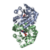
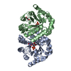
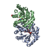
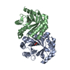
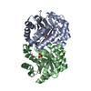
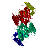
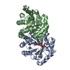
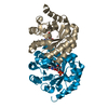
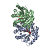
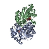
- PDB-1lp6: Crystal structure of orotidine monophosphate decarboxylase comple... -
+ Open data

- Basic information
| Entry | Database: PDB / ID: 1lp6 | ||||||
|---|---|---|---|---|---|---|---|
| Title | Crystal structure of orotidine monophosphate decarboxylase complexed with CMP | ||||||
 Components Components | orotidine monophosphate decarboxylase | ||||||
 Keywords Keywords |  LYASE / TIM barrel LYASE / TIM barrel | ||||||
| Function / homology |  Function and homology informationorotidine-5'-phosphate decarboxylase / orotidine-5'-phosphate decarboxylase activity / 'de novo' UMP biosynthetic process / 'de novo' pyrimidine nucleobase biosynthetic process Function and homology informationorotidine-5'-phosphate decarboxylase / orotidine-5'-phosphate decarboxylase activity / 'de novo' UMP biosynthetic process / 'de novo' pyrimidine nucleobase biosynthetic processSimilarity search - Function | ||||||
| Biological species | Archaea (unknown) | ||||||
| Method | X-RAY DIFFRACTION / SYNCHROTRON / MOLECULAR REPLACEMENT / Resolution: 1.9 Å | ||||||
 Authors Authors | Wu, N. / Pai, E.F. | ||||||
 Citation Citation | Journal: J.Biol.Chem. / Year: 2002 Title: Crystal structures of inhibitor complexes reveal an alternate binding mode in orotidine-5'-monophosphate decarboxylase. Authors: Wu, N. / Pai, E.F. | ||||||
| History |
| ||||||
| Remark 999 | sequence Authors state that although residues 1 and 1001 are MET and residues 101 and 1101 are Arg ... sequence Authors state that although residues 1 and 1001 are MET and residues 101 and 1101 are Arg according to the SwissProt entry, residues 1 and 1001 were LEU and residues 101 and 1101 were Pro in the original construct cloned of MT genomic dna. |
- Structure visualization
| Structure viewer | Molecule: MolmilJmol/JSmol |
|---|
- Downloads & links
-Download
| PDBx/mmCIF format | 1lp6.cif.gz | 95.8 KB | Display | PDBx/mmCIF format |
|---|---|---|---|---|
| PDB format | pdb1lp6.ent.gz | 72.7 KB | Display | PDB format |
| PDBx/mmJSON format | 1lp6.json.gz | Tree view | PDBx/mmJSON format | |
| Others |  Other downloads Other downloads |
-Validation report
| Arichive directory | https://data.pdbj.org/pub/pdb/validation_reports/lp/1lp6ftp://data.pdbj.org/pub/pdb/validation_reports/lp/1lp6 | HTTPS FTP |
|---|
-Related structure data


-Links
 PDBj
PDBj
- Assembly
| Deposited unit | 
| ||||||||
|---|---|---|---|---|---|---|---|---|---|
| 1 |
| ||||||||
| Unit cell |
| ||||||||
| Details | The biological unit is a dimer. |
-Components
| #1: Protein | Mass: 24994.830 Da / Num. of mol.: 2 Source method: isolated from a genetically manipulated source Source: (gene. exp.) Archaea (unknown) / Plasmid: pET15b / Production host:  Escherichia coli (E. coli) Escherichia coli (E. coli)References: UniProt: O26232, orotidine-5'-phosphate decarboxylase#2: Chemical | Cytidine monophosphate  Mass: 323.197 Da / Num. of mol.: 2 / Source method: obtained synthetically / Formula: C9H14N3O8P Mass: 323.197 Da / Num. of mol.: 2 / Source method: obtained synthetically / Formula: C9H14N3O8P#3: Water | ChemComp-HOH / | Water Mass: 18.015 Da / Num. of mol.: 162 / Source method: isolated from a natural source / Formula: H2O Mass: 18.015 Da / Num. of mol.: 162 / Source method: isolated from a natural source / Formula: H2O |
|---|
-Experimental details
-Experiment
| Experiment | Method: X-RAY DIFFRACTION / Number of used crystals: 1 |
|---|
- Sample preparation
| Crystal | Density Matthews: 2.09 Å3/Da / Density % sol: 41.04 % | ||||||||||||||||||||
|---|---|---|---|---|---|---|---|---|---|---|---|---|---|---|---|---|---|---|---|---|---|
| Crystal grow | Temperature: 298 K / Method: vapor diffusion, hanging drop / pH: 7.5 Details: trisodium citrate, 1,2,3-heptanetriol, 12-crown-4, pH 7.5, VAPOR DIFFUSION, HANGING DROP, temperature 25K | ||||||||||||||||||||
| Crystal grow | *PLUS | ||||||||||||||||||||
| Components of the solutions | *PLUS
|
-Data collection
| Diffraction | Mean temperature: 100 K |
|---|---|
| Diffraction source | Source: SYNCHROTRON / Site: APS  / Beamline: 14-BM-C / Wavelength: 1 Å / Beamline: 14-BM-C / Wavelength: 1 Å |
| Detector | Type: ADSC QUANTUM 4 / Detector: CCD / Date: Jun 15, 2001 |
| Radiation | Protocol: SINGLE WAVELENGTH / Monochromatic (M) / Laue (L): M / Scattering type: x-ray |
| Radiation wavelength | Wavelength: 1 Å / Relative weight: 1 |
| Reflection | Resolution: 1.9→30 Å / Num. all: 32661 / Num. obs: 32661 / % possible obs: 99.9 % / Observed criterion σ(F): 0 / Observed criterion σ(I): 0 / Biso Wilson estimate: 14.7 Å2 |
| Reflection shell | Resolution: 1.9→1.97 Å / % possible all: 99.9 |
| Reflection | *PLUS Lowest resolution: 30 Å / Num. measured all: 765425 / Rmerge(I) obs: 0.04 |
| Reflection shell | *PLUS % possible obs: 99.9 % / Rmerge(I) obs: 0.1 |
- Processing
| Software |
| ||||||||||||||||||||||||||||||||||||
|---|---|---|---|---|---|---|---|---|---|---|---|---|---|---|---|---|---|---|---|---|---|---|---|---|---|---|---|---|---|---|---|---|---|---|---|---|---|
| Refinement | Method to determine structure: MOLECULAR REPLACEMENT / Resolution: 1.9→28.47 Å / Rfactor Rfree error: 0.007 / Isotropic thermal model: RESTRAINED / Cross valid method: THROUGHOUT / σ(F): 0
| ||||||||||||||||||||||||||||||||||||
| Solvent computation | Solvent model: FLAT MODEL / Bsol: 48.158 Å2 / ksol: 0.372615 e/Å3 | ||||||||||||||||||||||||||||||||||||
| Displacement parameters | Biso mean: 27.9 Å2
| ||||||||||||||||||||||||||||||||||||
| Refine analyze | Luzzati coordinate error free: 0.26 Å / Luzzati sigma a free: 0.18 Å | ||||||||||||||||||||||||||||||||||||
| Refinement step | Cycle: LAST / Resolution: 1.9→28.47 Å
| ||||||||||||||||||||||||||||||||||||
| Refine LS restraints |
| ||||||||||||||||||||||||||||||||||||
| LS refinement shell | Resolution: 1.9→2.02 Å / Rfactor Rfree error: 0.018 / Total num. of bins used: 6
| ||||||||||||||||||||||||||||||||||||
| Xplor file |
| ||||||||||||||||||||||||||||||||||||
| Refinement | *PLUS Highest resolution: 1.9 Å / Lowest resolution: 30 Å / Rfactor all: 0.192 / Rfactor Rfree: 0.24 / Rfactor Rwork: 0.193 | ||||||||||||||||||||||||||||||||||||
| Solvent computation | *PLUS | ||||||||||||||||||||||||||||||||||||
| Displacement parameters | *PLUS | ||||||||||||||||||||||||||||||||||||
| Refine LS restraints | *PLUS
| ||||||||||||||||||||||||||||||||||||
| LS refinement shell | *PLUS Highest resolution: 1.9 Å / Rfactor Rfree: 0.272 / Rfactor Rwork: 0.215 |
