 Movie
Movie Controller

[English] 日本語
 Yorodumi
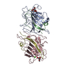
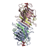
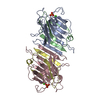
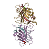
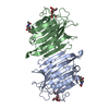
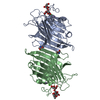
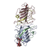
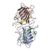
- PDB-1loa: THREE-DIMENSIONAL STRUCTURES OF COMPLEXES OF LATHYRUS OCHRUS ISOL... -
+ Open data

- Basic information
| Entry | Database: PDB / ID: 1loa | ||||||
|---|---|---|---|---|---|---|---|
| Title | THREE-DIMENSIONAL STRUCTURES OF COMPLEXES OF LATHYRUS OCHRUS ISOLECTIN I WITH GLUCOSE AND MANNOSE: FINE SPECIFICITY OF THE MONOSACCHARIDE-BINDING SITE | ||||||
 Components Components | (LEGUME ISOLECTIN I ...) x 2 | ||||||
 Keywords Keywords |  LECTIN LECTIN | ||||||
| Function / homology |  Function and homology information Function and homology information | ||||||
| Biological species |  Lathyrus ochrus (yellow-flowered pea) Lathyrus ochrus (yellow-flowered pea) | ||||||
| Method | X-RAY DIFFRACTION / Resolution: 2.2 Å | ||||||
 Authors Authors | Bourne, Y. / Cambillau, C. | ||||||
 Citation Citation | Journal: Proteins / Year: 1990 Title: Three-dimensional structures of complexes of Lathyrus ochrus isolectin I with glucose and mannose: fine specificity of the monosaccharide-binding site. Authors: Bourne, Y. / Roussel, A. / Frey, M. / Rouge, P. / Fontecilla-Camps, J.C. / Cambillau, C. #1: Journal: J.Mol.Biol. / Year: 1988Title: Crystallization and Preliminary X-Ray Studies of Two Isolectins from the Seeds of Lathyrus Ochrus Authors: Bourne, Y. / Rouge, P. / Cambillau, C. | ||||||
| History |
|
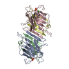
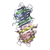
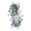
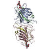
- Structure visualization
| Structure viewer | Molecule: MolmilJmol/JSmol |
|---|
- Downloads & links
-Download
| PDBx/mmCIF format | 1loa.cif.gz | 192.5 KB | Display | PDBx/mmCIF format |
|---|---|---|---|---|
| PDB format | pdb1loa.ent.gz | 153.7 KB | Display | PDB format |
| PDBx/mmJSON format | 1loa.json.gz | Tree view | PDBx/mmJSON format | |
| Others |  Other downloads Other downloads |
-Validation report
| Arichive directory | https://data.pdbj.org/pub/pdb/validation_reports/lo/1loaftp://data.pdbj.org/pub/pdb/validation_reports/lo/1loa | HTTPS FTP |
|---|
-Related structure data


-Links
 PDBj
PDBj

- Assembly
| Deposited unit | 
| ||||||||
|---|---|---|---|---|---|---|---|---|---|
| 1 | 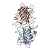
| ||||||||
| 2 | 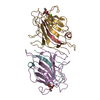
| ||||||||
| Unit cell |
| ||||||||
| Atom site foot note | 1: ALA A 80 - ASP A 81 OMEGA = 350.51 PEPTIDE BOND DEVIATES SIGNIFICANTLY FROM TRANS CONFORMATION 2: ALA C 80 - ASP C 81 OMEGA = 343.82 PEPTIDE BOND DEVIATES SIGNIFICANTLY FROM TRANS CONFORMATION 3: ALA E 80 - ASP E 81 OMEGA = 10.65 PEPTIDE BOND DEVIATES SIGNIFICANTLY FROM TRANS CONFORMATION 4: ALA G 80 - ASP G 81 OMEGA = 330.77 PEPTIDE BOND DEVIATES SIGNIFICANTLY FROM TRANS CONFORMATION | ||||||||
| Details | THIS CRYSTAL FORM CONTAINS TWO DIMERS IN THE ASYMMETRIC UNIT. EACH MONOMER CONSISTS OF TWO SEPARATE POLYPEPTIDE CHAINS, ALPHA AND BETA. THE ALPHA CHAIN CONSISTS OF 181 RESIDUES AND THE BETA CHAIN CONSISTS OF 52 RESIDUES. THE BETA AND ALPHA CHAINS OF MONOMER 1 HAVE BEEN ASSIGNED *A* AND *B*. THE BETA AND ALPHA CHAINS OF MONOMER 2 HAVE BEEN ASSIGNED *C* AND *D*. THE SAME ASSIGNMENT HAS BEEN MADE FOR THE SECOND MOLECULE WITH CHAIN IDENTIFIERS *E*, *F*, *G*, *H*. EACH DIMER HAS THE TWO MONOMERS WHICH ARE RELATED BY A NON-CRYSTALLOGRAPHIC TWO-FOLD AXIS. |
-Components
-LEGUME ISOLECTIN I ... , 2 types, 8 molecules ACEGBDFH
| #1: Protein | Mass: 19847.857 Da / Num. of mol.: 4 Source method: isolated from a genetically manipulated source Source: (gene. exp.) Lathyrus ochrus (yellow-flowered pea) / Organ: SEED / References: UniProt: P04122#2: Protein | Mass: 5783.322 Da / Num. of mol.: 4 Source method: isolated from a genetically manipulated source Source: (gene. exp.) Lathyrus ochrus (yellow-flowered pea) / Organ: SEED / References: UniProt: P12306 |
|---|
-Sugars , 1 types, 4 molecules 
| #3: Sugar | ChemComp-GYP / Methyl group Type: D-saccharide / Mass: 194.182 Da / Num. of mol.: 4 Type: D-saccharide / Mass: 194.182 Da / Num. of mol.: 4Source method: isolated from a genetically manipulated source Formula: C7H14O6 |
|---|
-Non-polymers , 3 types, 306 molecules 


| #4: Chemical | ChemComp-CA /  Mass: 40.078 Da / Num. of mol.: 4 / Source method: obtained synthetically / Formula: Ca Mass: 40.078 Da / Num. of mol.: 4 / Source method: obtained synthetically / Formula: Ca#5: Chemical | ChemComp-MN /  Mass: 54.938 Da / Num. of mol.: 4 / Source method: obtained synthetically / Formula: Mn Mass: 54.938 Da / Num. of mol.: 4 / Source method: obtained synthetically / Formula: Mn#6: Water | ChemComp-HOH / | WaterMass: 18.015 Da / Num. of mol.: 298 / Source method: isolated from a natural source / Formula: H2O |
|---|
-Details
| Sequence details | SEQUENCE ADVISORY NOTICE: DIFFERENCE BETWEEN SWISS-PROT AND PDB SEQUENCE. SWISS-PROT ENTRY NAME: ...SEQUENCE ADVISORY NOTICE: DIFFERENCE |
|---|
-Experimental details
-Experiment
| Experiment | Method: X-RAY DIFFRACTION |
|---|
- Sample preparation
| Crystal | Density Matthews: 2.41 Å3/Da / Density % sol: 48.87 % | ||||||||||||||||||||||||||||||
|---|---|---|---|---|---|---|---|---|---|---|---|---|---|---|---|---|---|---|---|---|---|---|---|---|---|---|---|---|---|---|---|
| Crystal grow | *PLUS Temperature: 20 ℃ / pH: 7.5 / Method: vapor diffusion, sitting dropDetails: reservoirs contain the same ingredients as drops, but twice as consentrated. taken from Bourne, Y. et al. (1988). J. Mol. Biol., 202, 685-687. | ||||||||||||||||||||||||||||||
| Components of the solutions | *PLUS
|
-Data collection
| Radiation | Scattering type: x-ray |
|---|---|
| Radiation wavelength | Relative weight: 1 |
| Reflection | *PLUS Highest resolution: 2.2 Å / Num. obs: 33640 / % possible obs: 67 % / Num. measured all: 77034 / Rmerge(I) obs: 0.082 |
- Processing
| Software |
| ||||||||||||||||||||||||||||||||||||||||||||||||||||||||||||
|---|---|---|---|---|---|---|---|---|---|---|---|---|---|---|---|---|---|---|---|---|---|---|---|---|---|---|---|---|---|---|---|---|---|---|---|---|---|---|---|---|---|---|---|---|---|---|---|---|---|---|---|---|---|---|---|---|---|---|---|---|---|
| Refinement | Resolution: 2.2→8 Å / Rfactor Rwork: 0.179 / Rfactor obs: 0.179 / σ(F): 1 | ||||||||||||||||||||||||||||||||||||||||||||||||||||||||||||
| Refinement step | Cycle: LAST / Resolution: 2.2→8 Å
| ||||||||||||||||||||||||||||||||||||||||||||||||||||||||||||
| Refine LS restraints |
| ||||||||||||||||||||||||||||||||||||||||||||||||||||||||||||
| Software | *PLUS Name: X-PLOR / Classification: refinement | ||||||||||||||||||||||||||||||||||||||||||||||||||||||||||||
| Refinement | *PLUS Rfactor obs: 0.179 / Rfactor Rwork: 0.179 | ||||||||||||||||||||||||||||||||||||||||||||||||||||||||||||
| Solvent computation | *PLUS | ||||||||||||||||||||||||||||||||||||||||||||||||||||||||||||
| Displacement parameters | *PLUS | ||||||||||||||||||||||||||||||||||||||||||||||||||||||||||||
| Refine LS restraints | *PLUS
|
