 Movie
Movie Controller


+ Open data

- Basic information
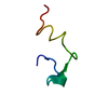
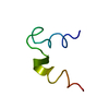
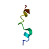
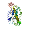
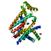
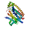
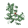
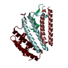
| Entry | Database: PDB / ID: 1ln6 | ||||||
|---|---|---|---|---|---|---|---|
| Title | STRUCTURE OF BOVINE RHODOPSIN (Metarhodopsin II) | ||||||
 Components Components | RHODOPSIN | ||||||
 Keywords Keywords | MEMBRANE PROTEIN / G-PROTEIN COUPLED RECEPTOR / METARHODOPSIN II / RHODOPSIN | ||||||
| Function / homology |  Function and homology informationOpsins / VxPx cargo-targeting to cilium / rod photoreceptor outer segment / rod bipolar cell differentiation / sperm head plasma membrane / podosome assembly / absorption of visible light / opsin binding / The canonical retinoid cycle in rods (twilight vision) / : ...Opsins / VxPx cargo-targeting to cilium / rod photoreceptor outer segment / rod bipolar cell differentiation / sperm head plasma membrane / podosome assembly / absorption of visible light / opsin binding / The canonical retinoid cycle in rods (twilight vision) / : / G protein-coupled photoreceptor activity / photoreceptor inner segment membrane / rhodopsin mediated signaling pathway / 11-cis retinal binding / cellular response to light stimulus / G protein-coupled receptor complex / Inactivation, recovery and regulation of the phototransduction cascade / phototransduction, visible light / thermotaxis / Activation of the phototransduction cascade / detection of temperature stimulus involved in thermoception / outer membrane / arrestin family protein binding / photoreceptor cell maintenance / photoreceptor outer segment membrane / G alpha (i) signalling events / response to light stimulus / phototransduction / photoreceptor outer segment / G-protein alpha-subunit binding / sperm midpiece / visual perception / guanyl-nucleotide exchange factor activity / microtubule cytoskeleton organization / photoreceptor disc membrane / cell-cell junction / gene expression / G protein-coupled receptor signaling pathway / Golgi membrane / zinc ion binding / membrane / identical protein binding / plasma membrane Function and homology informationOpsins / VxPx cargo-targeting to cilium / rod photoreceptor outer segment / rod bipolar cell differentiation / sperm head plasma membrane / podosome assembly / absorption of visible light / opsin binding / The canonical retinoid cycle in rods (twilight vision) / : ...Opsins / VxPx cargo-targeting to cilium / rod photoreceptor outer segment / rod bipolar cell differentiation / sperm head plasma membrane / podosome assembly / absorption of visible light / opsin binding / The canonical retinoid cycle in rods (twilight vision) / : / G protein-coupled photoreceptor activity / photoreceptor inner segment membrane / rhodopsin mediated signaling pathway / 11-cis retinal binding / cellular response to light stimulus / G protein-coupled receptor complex / Inactivation, recovery and regulation of the phototransduction cascade / phototransduction, visible light / thermotaxis / Activation of the phototransduction cascade / detection of temperature stimulus involved in thermoception / outer membrane / arrestin family protein binding / photoreceptor cell maintenance / photoreceptor outer segment membrane / G alpha (i) signalling events / response to light stimulus / phototransduction / photoreceptor outer segment / G-protein alpha-subunit binding / sperm midpiece / visual perception / guanyl-nucleotide exchange factor activity / microtubule cytoskeleton organization / photoreceptor disc membrane / cell-cell junction / gene expression / G protein-coupled receptor signaling pathway / Golgi membrane / zinc ion binding / membrane / identical protein binding / plasma membraneSimilarity search - Function | ||||||
| Method | SOLUTION NMR / MULTI-DIMENSIONAL NMR,CD, SIMULATED ANNEALING WITH MMFF94 FORCE FIELD, MOLECULAR DYNAMICS WITH CHARMM FORCE FIELD | ||||||
 Authors Authors | Choi, G. / Landin, J. / Galan, J.F. / Birge, R.R. / Albert, A.D. / Yeagle, P.L. | ||||||
 Citation Citation | Journal: Biochemistry / Year: 2002 Title: Structural studies of metarhodopsin II, the activated form of the G-protein coupled receptor, rhodopsin. Authors: Choi, G. / Landin, J. / Galan, J.F. / Birge, R.R. / Albert, A.D. / Yeagle, P.L. | ||||||
| History |
|
- Structure visualization
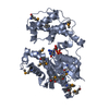
| Structure viewer | Molecule: MolmilJmol/JSmol |
|---|
- Downloads & links
-Download
| PDBx/mmCIF format | 1ln6.cif.gz | 102.8 KB | Display | PDBx/mmCIF format |
|---|---|---|---|---|
| PDB format | pdb1ln6.ent.gz | 80.5 KB | Display | PDB format |
| PDBx/mmJSON format | 1ln6.json.gz | Tree view | PDBx/mmJSON format | |
| Others |  Other downloads Other downloads |
-Validation report
| Arichive directory | https://data.pdbj.org/pub/pdb/validation_reports/ln/1ln6ftp://data.pdbj.org/pub/pdb/validation_reports/ln/1ln6 | HTTPS FTP |
|---|
-Related structure data
| Related structure data | |
|---|---|
| Similar structure data |


-Links
 PDBj
PDBj


- Assembly
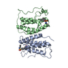
| Deposited unit | 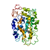
| |||||||||
|---|---|---|---|---|---|---|---|---|---|---|
| 1 |
| |||||||||
| NMR ensembles |
|
-Components
| #1: Protein | Mass: 39031.457 Da / Num. of mol.: 1 / Source method: obtained synthetically Details: This protein was chemically synthesized using a SOLID STATE PEPTIDE SYNTHESIZER. It is naturally found in Bos taurus (bovine) References: UniProt: P02699 |
|---|---|
| #2: Chemical | ChemComp-RET / Retinal  Mass: 284.436 Da / Num. of mol.: 1 / Source method: obtained synthetically / Formula: C20H28O Mass: 284.436 Da / Num. of mol.: 1 / Source method: obtained synthetically / Formula: C20H28O |
-Experimental details
-Experiment
| Experiment | Method: SOLUTION NMR | ||||||||||||
|---|---|---|---|---|---|---|---|---|---|---|---|---|---|
| NMR experiment |
|
- Sample preparation
| Details | Contents: FRAGMENTS OF RHODOPSIN / Solvent system: D2O, DMSO, DPC MICELLES |
|---|---|
| Sample conditions | Ionic strength: 10 mM / pH: 6.00 / Pressure: 1 atm / Temperature: 283.00 K |
| Crystal grow | *PLUS Method: other / Details: NMR |
-NMR measurement
| NMR spectrometer | Type: Bruker AMX / Manufacturer: Bruker / Model: AMX / Field strength: 600 MHz |
|---|
- Processing
| NMR software |
| ||||||||||||
|---|---|---|---|---|---|---|---|---|---|---|---|---|---|
| Refinement | Method: MULTI-DIMENSIONAL NMR,CD, SIMULATED ANNEALING WITH MMFF94 FORCE FIELD, MOLECULAR DYNAMICS WITH CHARMM FORCE FIELD Software ordinal: 1 Details: SECONDARY STRUCTURE FROM NMR STRUCTURES OF PEPTIDES ENCODING HELICES OR TURNS, AND LONG-RANGE DISTANCE CONSTRAINTS FROM OTHER MEASUREMENTS ON INTACT PROTEIN AMINO TERMINUS REMOVED DUE TO ...Details: SECONDARY STRUCTURE FROM NMR STRUCTURES OF PEPTIDES ENCODING HELICES OR TURNS, AND LONG-RANGE DISTANCE CONSTRAINTS FROM OTHER MEASUREMENTS ON INTACT PROTEIN AMINO TERMINUS REMOVED DUE TO LACK OF LONG-RANGE CONSTRAINTS | ||||||||||||
| NMR ensemble | Conformer selection criteria: structures with the lowest energy Conformers calculated total number: 10 / Conformers submitted total number: 1 |
