 Movie
Movie Controller


+ Open data

- Basic information
| Entry | Database: PDB / ID: 1lk9 | |||||||||
|---|---|---|---|---|---|---|---|---|---|---|
| Title | The Three-dimensional Structure of Alliinase from Garlic | |||||||||
 Components Components | ALLIIN LYASE Alliinase Alliinase | |||||||||
 Keywords Keywords | LYASE / EGF-like domain / PLP type 1 / chloride binding | |||||||||
| Function / homology |  Function and homology informationalliin lyase / alliin lyase activity / vacuole / chloride ion binding / pyridoxal phosphate binding / protein homodimerization activity Function and homology informationalliin lyase / alliin lyase activity / vacuole / chloride ion binding / pyridoxal phosphate binding / protein homodimerization activitySimilarity search - Function | |||||||||
| Biological species |  Allium sativum (garlic) Allium sativum (garlic) | |||||||||
| Method | X-RAY DIFFRACTION / SYNCHROTRON / SIRAS / Resolution: 1.53 Å | |||||||||
 Authors Authors | Kuettner, E.B. / Hilgenfeld, R. / Weiss, M.S. | |||||||||
 Citation Citation | Journal: J.Biol.Chem. / Year: 2002 Title: The active principle of garlic at atomic resolution Authors: Kuettner, E.B. / Hilgenfeld, R. / Weiss, M.S. #1: Journal: Arch.Biochem.Biophys. / Year: 2002Title: PURIFICATION, CHARACTERIZATION AND CRYSTALLIZATION OF ALLIINASE FROM GARLIC Authors: KUETTNER, E.B. / HILGENFELD, R. / WEISS, M.S. #2: Journal: Arch.Biochem.Biophys. / Year: 2002Title: Erratum to ``Purification, characterization and crystallization of alliinase from garlic. [Arch. Biochem. Biophys. 402, 192-200.]'' Authors: Kuettner, E.B. / Hilgenfeld, R. / Weiss, M.S. | |||||||||
| History |
| |||||||||
| Remark 600 | heterogen The molecules EPE and DHA are only present at an occupancy of 50%. Their presence is ...heterogen The molecules EPE and DHA are only present at an occupancy of 50%. Their presence is mutually exclusive. | |||||||||
| Remark 999 | sequence there are two different sequences in the literature, one contains an Asp in position 176 ...sequence there are two different sequences in the literature, one contains an Asp in position 176 and one contains Asn. The authors have built and refined the structure with an Asp present in this position. |
- Structure visualization
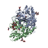
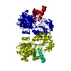
| Structure viewer | Molecule: MolmilJmol/JSmol |
|---|
- Downloads & links
-Download
| PDBx/mmCIF format | 1lk9.cif.gz | 204.1 KB | Display | PDBx/mmCIF format |
|---|---|---|---|---|
| PDB format | pdb1lk9.ent.gz | 165.3 KB | Display | PDB format |
| PDBx/mmJSON format | 1lk9.json.gz | Tree view | PDBx/mmJSON format | |
| Others |  Other downloads Other downloads |
-Validation report
| Arichive directory | https://data.pdbj.org/pub/pdb/validation_reports/lk/1lk9ftp://data.pdbj.org/pub/pdb/validation_reports/lk/1lk9 | HTTPS FTP |
|---|
-Related structure data
| Similar structure data |
|---|


-Links
 PDBj
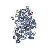
PDBj- Assembly
| Deposited unit | 
| ||||||||
|---|---|---|---|---|---|---|---|---|---|
| 1 |
| ||||||||
| Unit cell |
|
-Components
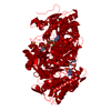
-Protein , 1 types, 2 molecules AB
| #1: Protein | Alliinase / ALLIINASE / CYSTEINE SULPHOXIDE LYASE Mass: 51516.477 Da / Num. of mol.: 2 / Source method: isolated from a natural source / Details: bulb / Source: (natural) Allium sativum (garlic) / References: UniProt: Q01594, alliin lyase |
|---|
-Sugars , 3 types, 4 molecules
| #2: Polysaccharide | beta-D-mannopyranose-(1-4)-2-acetamido-2-deoxy-beta-D-glucopyranose-(1-4)-[alpha-L-fucopyranose-(1- ...beta-D-mannopyranose-(1-4)-2-acetamido-2-deoxy-beta-D-glucopyranose-(1-4)-[alpha-L-fucopyranose-(1-3)]2-acetamido-2-deoxy-beta-D-glucopyranose / Mass: 732.682 Da / Num. of mol.: 1 Source method: isolated from a genetically manipulated source | ||
|---|---|---|---|
| #3: Polysaccharide | / Mass: 424.401 Da / Num. of mol.: 2 Source method: isolated from a genetically manipulated source #4: Polysaccharide | alpha-L-fucopyranose-(1-3)-2-acetamido-2-deoxy-beta-D-glucopyranose | / Mass: 367.349 Da / Num. of mol.: 1Source method: isolated from a genetically manipulated source |
-Non-polymers , 6 types, 848 molecules 


| #5: Chemical | ChemComp-SO4 / Sulfate Mass: 96.063 Da / Num. of mol.: 11 / Source method: obtained synthetically / Formula: SO4 Mass: 96.063 Da / Num. of mol.: 11 / Source method: obtained synthetically / Formula: SO4#6: Chemical | Chloride Mass: 35.453 Da / Num. of mol.: 2 / Source method: obtained synthetically / Formula: Cl Mass: 35.453 Da / Num. of mol.: 2 / Source method: obtained synthetically / Formula: Cl#7: Chemical | Pyridoxal phosphate Mass: 247.142 Da / Num. of mol.: 2 / Source method: obtained synthetically / Formula: C8H10NO6P Mass: 247.142 Da / Num. of mol.: 2 / Source method: obtained synthetically / Formula: C8H10NO6P#8: Chemical | HEPES Mass: 238.305 Da / Num. of mol.: 2 / Source method: obtained synthetically / Formula: C8H18N2O4S / Comment: pH buffer*YM Mass: 238.305 Da / Num. of mol.: 2 / Source method: obtained synthetically / Formula: C8H18N2O4S / Comment: pH buffer*YM#9: Chemical | ChemComp-DHA / | Dehydroalanine Type: peptide linking / Mass: 87.077 Da / Num. of mol.: 1 / Source method: obtained synthetically / Formula: C3H5NO2 Type: peptide linking / Mass: 87.077 Da / Num. of mol.: 1 / Source method: obtained synthetically / Formula: C3H5NO2#10: Water | ChemComp-HOH / | WaterMass: 18.015 Da / Num. of mol.: 830 / Source method: isolated from a natural source / Formula: H2O |
|---|
-Experimental details
-Experiment
| Experiment | Method: X-RAY DIFFRACTION / Number of used crystals: 1 |
|---|
- Sample preparation
| Crystal | Density Matthews: 2.49 Å3/Da / Density % sol: 50.6 % | ||||||||||||||||||||||||||||||||||||||||||
|---|---|---|---|---|---|---|---|---|---|---|---|---|---|---|---|---|---|---|---|---|---|---|---|---|---|---|---|---|---|---|---|---|---|---|---|---|---|---|---|---|---|---|---|
| Crystal grow | Temperature: 293 K / Method: vapor diffusion, hanging drop / pH: 7.4 Details: PROTEIN: 5 MG/ML ALLIINASE, 5 MM HEPES PH 7.4, 10 % (V/V) GLYCEROL, 0.25 MM PYRIDOXAL-5'-PHOSPHATE, 1 MM S-ETHYL-L-CYSTEINE; PRECIPITANT: 2.9 M AMMONIUM SULFATE, 50 MM HEPES PH 7.4 HANGING ...Details: PROTEIN: 5 MG/ML ALLIINASE, 5 MM HEPES PH 7.4, 10 % (V/V) GLYCEROL, 0.25 MM PYRIDOXAL-5'-PHOSPHATE, 1 MM S-ETHYL-L-CYSTEINE; PRECIPITANT: 2.9 M AMMONIUM SULFATE, 50 MM HEPES PH 7.4 HANGING DROP METHOD(4 MICROL + 4 MICROL), VAPOUR DIFFUSION, VAPOR DIFFUSION, HANGING DROP, temperature 293K | ||||||||||||||||||||||||||||||||||||||||||
| Crystal grow | *PLUS Temperature: 4 ℃Details: Kuettner, E.B., (2002) Arch.Biochem.Biophys., 402, 192. | ||||||||||||||||||||||||||||||||||||||||||
| Components of the solutions | *PLUS
|
-Data collection
| Diffraction | Mean temperature: 100 K |
|---|---|
| Diffraction source | Source: SYNCHROTRON / Site: EMBL/DESY, HAMBURG  / Beamline: BW7B / Wavelength: 0.8423 Å / Beamline: BW7B / Wavelength: 0.8423 Å |
| Detector | Type: MARRESEARCH / Detector: IMAGE PLATE / Date: Apr 25, 2000 |
| Radiation | Protocol: SINGLE WAVELENGTH / Monochromatic (M) / Laue (L): M / Scattering type: x-ray |
| Radiation wavelength | Wavelength: 0.8423 Å / Relative weight: 1 |
| Reflection | Resolution: 1.53→12 Å / Num. all: 160747 / Num. obs: 160747 / % possible obs: 98.9 % / Observed criterion σ(F): 0 / Observed criterion σ(I): -3 / Redundancy: 5.8 % / Biso Wilson estimate: 20.7 Å2 / Rmerge(I) obs: 0.055 / Net I/σ(I): 20.3 |
| Reflection shell | Resolution: 1.53→1.56 Å / Redundancy: 3.7 % / Rmerge(I) obs: 0.508 / Mean I/σ(I) obs: 2.1 / Num. unique all: 8024 / % possible all: 99.9 |
| Reflection | *PLUS Lowest resolution: 20 Å / Num. measured all: 928820 / Rmerge(I) obs: 0.057 |
| Reflection shell | *PLUS % possible obs: 99.6 % |
- Processing
| Software |
| ||||||||||||||||||||||||||||||||||||||||||||||||||||||||||||
|---|---|---|---|---|---|---|---|---|---|---|---|---|---|---|---|---|---|---|---|---|---|---|---|---|---|---|---|---|---|---|---|---|---|---|---|---|---|---|---|---|---|---|---|---|---|---|---|---|---|---|---|---|---|---|---|---|---|---|---|---|---|
| Refinement | Method to determine structure: SIRAS / Resolution: 1.53→12 Å / SU B: 1.52975 / SU ML: 0.05519 / Isotropic thermal model: isotropic / Cross valid method: THROUGHOUT / σ(F): 0 / ESU R: 0.07531 / ESU R Free: 0.07692 / Stereochemistry target values: Engh & Huber
| ||||||||||||||||||||||||||||||||||||||||||||||||||||||||||||
| Displacement parameters | Biso mean: 25.8 Å2 | ||||||||||||||||||||||||||||||||||||||||||||||||||||||||||||
| Refine analyze |
| ||||||||||||||||||||||||||||||||||||||||||||||||||||||||||||
| Refinement step | Cycle: LAST / Resolution: 1.53→12 Å
| ||||||||||||||||||||||||||||||||||||||||||||||||||||||||||||
| Refine LS restraints |
| ||||||||||||||||||||||||||||||||||||||||||||||||||||||||||||
| LS refinement shell | Resolution: 1.53→1.604 Å
| ||||||||||||||||||||||||||||||||||||||||||||||||||||||||||||
| Refinement | *PLUS Lowest resolution: 20 Å / Rfactor Rfree: 0.221 / Rfactor Rwork: 0.193 | ||||||||||||||||||||||||||||||||||||||||||||||||||||||||||||
| Solvent computation | *PLUS | ||||||||||||||||||||||||||||||||||||||||||||||||||||||||||||
| Displacement parameters | *PLUS | ||||||||||||||||||||||||||||||||||||||||||||||||||||||||||||
| Refine LS restraints | *PLUS
|
